





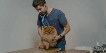
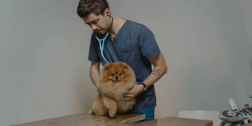
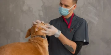







Quick take: Optic nerve swelling in dogs, also called optic nerve edema, is a condition where the nerve that carries visual information from the eye to the brain becomes inflamed or enlarged. It can cause vision loss, pain, and, depending on the cause, may be urgent. Prompt veterinary evaluation, imaging, and targeted treatment give the best chance for visual recovery and a good quality of life.
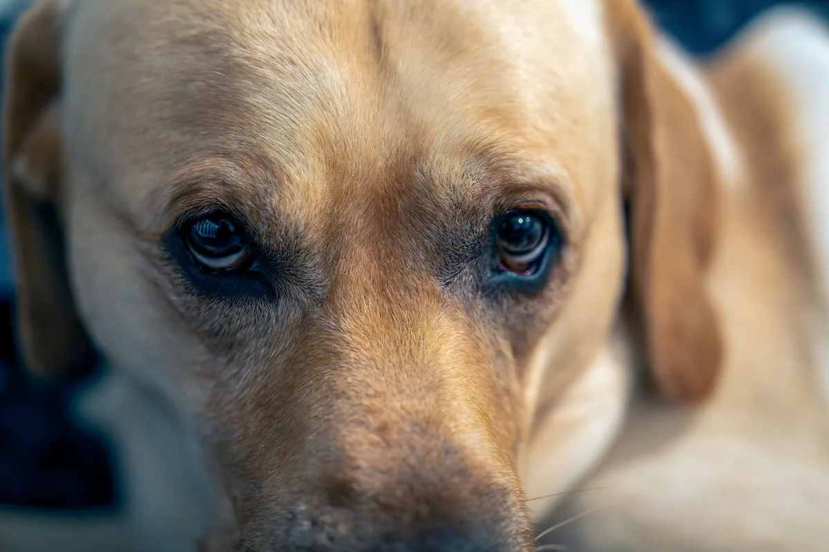
It’s 11 p.m., and your 7‑year‑old mixed‑breed Labrador is lying on the couch, eyes half‑closed, and seems unusually sluggish. When you gently lift his head, you notice his right eye looks a bit cloudier than usual, and his pupils don’t respond the way they normally do. Your heart starts racing and you type “why won’t my dog see” into the search bar. You’re not alone—many owners discover optic nerve swelling after a subtle change in their dog’s behavior or eye appearance.
Optic nerve swelling (optic nerve edema) is a serious, but often treatable, eye problem. The swelling itself is a symptom of an underlying issue, such as inflammation, a tumor, trauma, infection, or a systemic disease. Your vet will need to pinpoint the cause before deciding on the right therapy.
In this article we’ll explain what the optic nerve is, why it can swell, what signs to watch for, how vets diagnose it, treatment options (medical and surgical), diet considerations, typical costs, and how you can help prevent future problems. We’ll also give you a clear action plan for when you should call your vet right away.
What is optic nerve swelling in dogs?
Optic nerve swelling, medically termed optic nerve edema, is the enlargement of the optic nerve—a bundle of nerve fibers that transmits visual signals from the retina to the brain. In dogs the optic nerve travels from the back of the eye, through the optic canal, and into the brain’s visual centers. When the nerve becomes inflamed or filled with fluid, its shape changes, and pressure can disrupt signal transmission, leading to visual deficits or blindness.
While any breed can develop optic nerve swelling, it is relatively uncommon, representing a small fraction of ophthalmic referrals in veterinary hospitals. According to the American College of Veterinary Ophthalmologists (ACVO), optic nerve pathology accounts for roughly 2–3 % of all eye‑related cases seen in specialty practices.
What causes optic nerve swelling in dogs?
Swelling of the optic nerve can arise from several distinct pathways. Understanding the underlying cause guides treatment and prognosis.
- Inflammatory (optic neuritis): Autoimmune disorders, such as granulomatous meningoencephalitis, or infectious agents (e.g., canine distemper virus, Toxoplasma gondii) trigger inflammation of the nerve.
- Neoplastic (tumors): Primary optic nerve gliomas or metastatic tumors (e.g., lymphoma, meningioma) can compress or infiltrate the nerve.
- Trauma: Blunt force, penetrating injuries, or severe head trauma can cause hemorrhage or edema within the optic nerve.
- Infectious or parasitic: Fungal infections (Cryptococcus), bacterial meningitis, or heartworm disease can extend inflammation to the optic nerve.
- Systemic disease: Hypertension, immune‑mediated diseases, or severe anemia can lead to secondary optic nerve swelling.
Signs and symptoms
Because the optic nerve lies deep inside the skull, owners often notice changes in vision or eye appearance before swelling is obvious on imaging. Early signs can be subtle, while severe cases present dramatic visual loss.
| Severity | Typical signs |
|---|---|
| Mild | Reluctance to chase moving objects, occasional bumping into furniture, slight pupil asymmetry. |
| Moderate | Noticeable cloudiness of the eye, reduced response to light, hesitancy to go up or down stairs. |
| Severe | Complete blindness in one or both eyes, eye pain (blepharospasm), dilated non‑reactive pupil, head tilt, loss of balance. |
When to call your vet
Call your vet today if you notice any of the following:
- Sudden cloudiness or redness in one eye.
- Pupil that does not constrict to light.
- Loss of interest in fetch, climbing stairs, or navigating familiar spaces.
- Head tilt or unsteady gait.
Go to an emergency veterinary hospital right now if you see:
- Severe eye pain (excessive squinting, rubbing the eye).
- Sudden, complete blindness in both eyes.
- Bleeding from the eye or obvious trauma.
These signs can indicate rapid progression or an underlying life‑threatening condition. This article provides general information and is not a substitute for professional veterinary care.
How vets diagnose optic nerve swelling in dogs
Diagnosing optic nerve swelling involves a stepwise workup that combines a thorough history, physical exam, and specialized tests.
- Ophthalmic examination: Using an ophthalmoscope, the vet looks at the optic disc for swelling (papilledema) and assesses pupil reflexes.
- Fundus photography: Captures images of the retina and optic nerve for comparison over time.
- Imaging: MRI is the gold standard for visualizing nerve enlargement and differentiating causes (tumor vs. inflammation). CT can be used when MRI is unavailable.
- Neurologic exam: Checks for other signs of brain involvement (e.g., seizures, altered mentation).
- Laboratory tests: CBC, chemistry panel, and infectious disease titers (e.g., distemper, Toxoplasma) help identify systemic contributors.
Treatment options
Therapy is tailored to the underlying cause and the severity of swelling.
Medical treatment
When inflammation or infection is the primary driver, most dogs benefit from anti‑inflammatory and antimicrobial medications. Common drug classes include:
- Corticosteroids: Prednisone or dexamethasone to reduce immune‑mediated swelling (ask your vet about this option).
- Immunosuppressants: Cyclosporine or mycophenolate mofetil for autoimmune optic neuritis (vet‑guided dosing).
- Antibiotics/antifungals: Enrofloxacin for bacterial meningitis, fluconazole for fungal infections (vet will choose based on culture results).
- Antiviral agents: Acyclovir may be used for severe canine distemper‑related neuritis (if indicated).
Supplements and supportive care
Adjunctive nutrition can aid recovery, especially when inflammation is present:
- Omega‑3 fatty acids (EPA/DHA): Reduce ocular inflammation; 300–500 mg per day of a high‑quality fish‑oil supplement is typical for a medium‑size dog (ask your vet).
- Antioxidants: Vitamin E and lutein support retinal health; many commercial “eye health” supplements contain these ingredients.
- Probiotics: Helpful if systemic infection or antibiotic therapy disrupts gut flora.
Supplements should complement, not replace, prescribed medication.
Procedures or surgery
If a tumor or severe structural compression is identified, surgical intervention may be necessary. Options include:
- Optic nerve sheath decompression: Rarely performed, aims to relieve pressure.
- Neuro‑oncologic surgery: Resection of accessible optic gliomas or meningiomas, often combined with radiation therapy.
- Radiation therapy: Stereotactic radiosurgery can shrink tumors without invasive surgery.
Recovery from optic nerve surgery varies; most dogs need 2–4 weeks of restricted activity and close neurologic monitoring. The decision for surgery is made after imaging and discussion of prognosis with your vet.
Diet and nutrition
While no single diet can cure optic nerve swelling, feeding a balanced, high‑quality diet supports overall neurologic health and can reduce inflammation. Here are evidence‑based guidelines for dogs dealing with optic nerve issues.
Foods to favor
- Highly digestible protein sources: Chicken, turkey, or salmon provide essential amino acids without taxing the liver.
- Omega‑3 rich ingredients: Fish oil, flaxseed, or chia seeds supply EPA/DHA, which have anti‑inflammatory effects on ocular tissues.
- Antioxidant‑rich fruits and vegetables: Blueberries, carrots, and spinach deliver lutein, zeaxanthin, and vitamin E, all of which protect retinal cells.
- Low‑sodium, moderate‑fat formulas: Particularly important if your dog has concurrent hypertension or systemic disease.
Foods to limit or avoid
- High‑fat, low‑quality fillers: Corn, wheat, and soy can promote systemic inflammation.
- Excessive vitamin A: Over‑supplementation can be toxic to the retina; stick to balanced commercial diets.
- Artificial additives: Preservatives and colors may trigger sensitivities in some dogs.
Many veterinary specialists recommend a therapeutic “neurological” diet when optic nerve swelling is linked to systemic disease. These diets are formulated to provide optimal levels of omega‑3s, antioxidants, and highly digestible proteins. Brands vary by region, but the nutrient profile is consistent with AAHA nutrition guidelines.
When transitioning to a new diet, do it gradually over 7–10 days to avoid gastrointestinal upset. Start with 25 % new food mixed with 75 % old, then increase the new food proportion every two days. Monitor your dog’s appetite, stool quality, and energy levels throughout the switch.
| Do feed | Limit | Avoid |
|---|---|---|
| High‑quality protein (chicken, salmon) | Moderate‑fat treats | Grain‑heavy filler diets |
| Omega‑3 sources (fish oil) | Excessive vitamin A supplements | Artificial colors/preservatives |
| Antioxidant‑rich veggies (blueberries) | High‑salt snacks | Raw bones with high fat |
Always discuss any diet change with your veterinarian, especially if your dog is on medication that could interact with certain nutrients (e.g., corticosteroids and calcium). Staying hydrated is also crucial; fresh water should be available at all times.
Cost and prognosis
Financial considerations can be a source of anxiety, so here’s a realistic breakdown of what you might expect.
| Service | US estimate | UK estimate |
|---|---|---|
| Comprehensive ophthalmic exam (including fundus photography) | $150–$250 | £80–£130 |
| MRI of the brain/orbit | $1,200–$2,200 | £700–£1,200 |
| Blood work & infectious disease panel | $200–$400 | £100–£180 |
| Medical therapy (3‑month course) | $300–$800 | £150–£400 |
| Surgical resection or radiation (if needed) | $3,000–$7,500 | £2,000–£5,000 |
Prognosis hinges on the underlying cause:
- Inflammatory optic neuritis: With prompt steroid therapy, 70–80 % of dogs regain functional vision within weeks.
- Neoplastic causes: Surgical removal or radiation can extend survival, but visual recovery is variable (30–50 % retain partial vision).
- Traumatic injury: Outcomes depend on severity; mild trauma often resolves, while severe nerve damage may be permanent.
Overall, early detection and treatment give the best odds for preserving sight and quality of life.
Prevention and home care
While you can’t control every cause of optic nerve swelling, several everyday practices reduce risk:
- Vaccinate: Keep your dog up‑to‑date on core vaccines (distemper, canine adenovirus) to lower infectious triggers.
- Control parasites: Monthly heartworm and flea/tick preventives (e.g., ivermectin‑based products) reduce the chance of systemic infections that could affect the optic nerve.
- Regular veterinary check‑ups: Annual eye exams catch subtle changes before they progress.
- Monitor blood pressure: Senior dogs benefit from routine blood pressure screening, as hypertension can cause papilledema.
- Safe environment: Prevent head injuries by avoiding rough play that could lead to trauma, especially in active breeds.
After treatment, follow your vet’s schedule for re‑checks—typically at 2 weeks, 1 month, and then every 3–6 months. Keep a journal of any visual changes, appetite, or behavior to share at each visit.
From our vet team: “Optic nerve swelling can feel overwhelming, but remember that many dogs recover vision when the underlying cause is addressed quickly. Keep a close eye on any changes, act fast if vision worsens, and lean on your vet for a tailored plan—early intervention is the key to a brighter outlook.”
Key takeaways
- Optic nerve swelling is an emergency‑level eye problem that can cause vision loss; prompt veterinary assessment is essential.
- Common causes include inflammation, tumors, trauma, infection, and systemic diseases such as hypertension.
- Watch for cloudiness, pupil changes, head tilt, or sudden blindness—these signs merit a vet call today or an ER visit if severe.
- Diagnosis relies on a thorough eye exam, MRI imaging, and blood work to uncover the root cause.
- Treatment may involve steroids, immunosuppressants, antibiotics, or surgery, depending on the cause.
- Feeding a high‑quality diet rich in omega‑3s and antioxidants supports recovery, while avoiding filler‑heavy, high‑salt foods helps reduce inflammation.
Myth vs. fact
Myth: Optic nerve swelling always leads to permanent blindness.
Fact: When caught early and treated appropriately, many dogs regain functional vision, especially if the swelling is due to inflammation rather than irreversible nerve damage.
Myth: Only older dogs develop optic nerve swelling.
Fact: While senior dogs are at higher risk for tumors and hypertension, young dogs can experience swelling from infections, trauma, or autoimmune disease.
Myth: Surgery is the only cure.
Fact: Medical management with steroids, immunosuppressants, or antibiotics can resolve swelling in many cases; surgery is reserved for tumors or refractory compression.
Frequently asked questions
What are the early signs of optic nerve swelling in a dog?
Early signs include subtle cloudiness of the eye, a pupil that doesn’t react to light, and the dog seeming less interested in chasing balls or navigating stairs. These changes often appear before any obvious blindness.
How do vets confirm optic nerve swelling in dogs?
Vets confirm swelling by performing a detailed ophthalmic exam and then imaging the brain and orbit with MRI, which visualizes the optic nerve’s size and any surrounding lesions.
Can medication shrink a swollen optic nerve in dogs?
Yes—anti‑inflammatory drugs such as corticosteroids and targeted immunosuppressants can reduce swelling when the cause is inflammatory or autoimmune. Antibiotics or antifungals work when an infection is responsible.
Will my dog lose its eyesight permanently from optic nerve swelling?
Permanent vision loss is possible if the nerve is severely damaged or if a tumor cannot be removed, but many dogs recover sight when treatment starts early and the underlying cause is addressed.
Is surgery required for optic nerve swelling in dogs?
Surgery is not always required; it is typically reserved for cases where a tumor or structural compression is identified and cannot be managed medically.
How much should I expect to pay for treatment of optic nerve swelling?
Overall costs range from a few hundred dollars for diagnostics and medication to several thousand dollars if advanced imaging or surgery is needed. See the cost table above for detailed estimates in the US and UK.
Ask the PuppaDogs community
Have a question this article didn’t fully answer? Want to compare notes with other dog owners who’ve been through this? Our community forum is moderated by experienced owners and vets — and answers tend to come fast. Ask in the PuppaDogs community →
References
- American College of Veterinary Ophthalmologists (ACVO). “Optic Nerve Disorders in Dogs.” Clinical guidelines, 2023.
- American Animal Hospital Association (AAHA). “Ophthalmic Care Standards.” 2022 update.
- Merck Veterinary Manual. “Optic Neuritis and Optic Nerve Edema.” Chapter 23, 2021.
- American College of Veterinary Internal Medicine (ACVIM). “Systemic Hypertension in Dogs.” Consensus statement, 2022.
- Veterinary Ophthalmology, 5th Edition. Gelatt, K.N., et al., Elsevier, 2020.
- University of California, Davis Veterinary Medicine. “MRI Findings in Canine Optic Nerve Tumors.” Veterinary Radiology Review, 2021.
- World Small Animal Veterinary Association (WSAVA). “Nutrition Guidelines for Dogs with Neurologic Disease.” 2023.


