








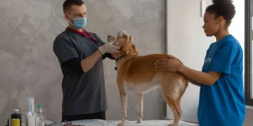
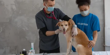






Quick take: A liver fistula is an abnormal tunnel that connects the liver to another organ, most often the gallbladder or intestine. It can cause vomiting, loss of appetite, and abdominal pain, and may be life‑threatening if left untreated. Prompt veterinary evaluation, imaging, and either medical management or surgery give most dogs a good chance at recovery.
It’s 11 p.m., and your 7‑year‑old mixed‑breed retriever, Max, is lying on the couch, eyes half‑closed, and refuses his usual bowl of kibble. When you check his gums, they look a little yellow‑green, and he lets out a soft whimper when you gently press his abdomen. Your mind races: “Is this something I can fix at home? Is it serious?” You’re not alone—many owners first notice a liver fistula when a normally energetic dog suddenly slows down, loses appetite, or shows subtle changes in stool and urine.
We understand how unsettling an unfamiliar diagnosis can feel. In this article we’ll explain exactly what a liver fistula is, why it occurs, what signs to watch for, how vets confirm the problem, and the range of treatment options—from medication to surgery. We’ll also cover diet tips, expected costs, recovery timelines, and steps you can take to lower the risk for your furry companion. By the end you’ll have a clear plan of action and know exactly when to call your vet.
What is a liver fistula in dogs?
A liver fistula is an abnormal channel—called a fistula—that forms between the liver and another nearby organ, most commonly the gallbladder (biliary fistula) or the intestine (enteric fistula). In a healthy dog, the liver processes nutrients, detoxifies waste, and produces bile that drains into the gallbladder. A fistula bypasses this normal route, allowing bile or intestinal contents to leak directly into the liver or vice‑versa. This can irritate liver tissue, cause inflammation, and lead to infection.
While liver fistulas are relatively rare, they are reported in up to 0.05 % of all canine necropsies, according to the American College of Veterinary Internal Medicine (ACVIM). Certain breeds—such as Cairn Terriers, Miniature Schnauzers, and Shih Tzus—appear predisposed, likely due to inherited anatomical quirks. Most dogs are diagnosed between 5 and 10 years of age, but congenital (present at birth) cases can be seen in puppies.
What causes it?
Fistulas can be congenital or acquired. The major categories are:
| Cause | Typical Mechanism |
|---|---|
| Congenital malformation | Abnormal development of bile ducts or liver tissue in utero, leading to a pre‑existing tunnel. |
| Chronic inflammation | Long‑standing hepatitis, cholecystitis, or pancreatitis erodes tissue and creates a passage. |
| Trauma or surgery | Blunt abdominal injury or previous liver surgery can inadvertently form a fistulous tract. |
| Infectious disease | Severe bacterial or parasitic infections (e.g., Leptospira) can damage liver walls. |
| Neoplasia | Cancerous growths may invade and perforate liver tissue. |
Breed predisposition is strongest for Cairn Terriers, who have a genetic tendency toward biliary tract anomalies. Miniature Schnauzers and Shih Tzus also show higher rates, possibly linked to their small size and unique biliary anatomy.
Signs and symptoms
Because the liver plays a central role in digestion, a fistula often manifests as gastrointestinal upset and systemic signs. Early clues can be subtle.
| Severity | Typical signs |
|---|---|
| Mild | Decreased appetite, occasional vomiting, slight weight loss, dull coat. |
| Moderate | Frequent vomiting (especially after meals), abdominal pain on palpation, yellow‑green gums, foul‑smelling stool. |
| Severe | Persistent vomiting, lethargy, jaundice (yellow skin), ascites (fluid in abdomen), fever, shock. |
Other red‑flag signs include a sudden change in urination frequency, blood‑tinged vomit, or obvious pain when you press on the right side of the abdomen. Some owners first notice a “bubbly” or “gritty” sound when the dog coughs, reflecting bile leakage into the chest cavity.
When to call your vet
Call your regular vet today if:
- Your dog shows mild to moderate vomiting, loss of appetite, or a change in stool color.
- You notice yellow‑green gums or a slight jaundice of the eyes.
- There’s any abdominal tenderness when you gently press the right flank.
Go to an emergency veterinary hospital right now if you see any of these signs:
- Persistent vomiting (more than two episodes in an hour) or vomiting blood.
- Severe lethargy, collapse, or inability to stand.
- Rapidly swelling abdomen (ascites) or signs of shock (rapid heartbeat, pale gums).
These recommendations are for information only and do not replace a hands‑on veterinary exam. If you’re ever unsure, err on the side of caution and seek professional care.
How vets diagnose it
Diagnosing a liver fistula involves a stepwise workup:
- History and physical exam: The vet will ask about diet, vomiting frequency, and any prior surgeries or illnesses, then palpate the abdomen for pain or fluid.
- Blood work: A complete blood count (CBC) and chemistry panel can reveal anemia, elevated liver enzymes (ALT, ALP), and bilirubin levels that point to liver dysfunction.
- Abdominal ultrasound: This is the most common imaging tool. Ultrasound can visualize an abnormal tract, fluid collections, or thickened bile ducts.
- CT scan or MRI (advanced): When ultrasound is inconclusive, cross‑sectional imaging provides a detailed map of the fistula’s course.
- Fistulography: A contrast dye is injected into the suspected tract under fluoroscopy, highlighting the connection on X‑ray.
- Liver biopsy: In some cases, a small sample of liver tissue is taken to assess inflammation or rule out cancer.
These tests together allow the veterinarian to differentiate a liver fistula from other liver diseases, such as a hepatic shunt or cholangitis.
Treatment options
Medical treatment
When surgery isn’t immediately feasible—or while awaiting an operation—vets may use medication to control inflammation, infection, and bile leakage. Common drug classes include:
- Broad‑spectrum antibiotics (e.g., amoxicillin‑clavulanate) to prevent secondary bacterial infection.
- Anti‑inflammatory drugs such as corticosteroids (prednisone) or non‑steroidal anti‑inflammatories (carprofen) to reduce liver swelling.
- Ursodeoxycholic acid (UDCA) to improve bile flow and protect liver cells.
- Antiemetics like maropitant (Cerenia) to control vomiting.
Ask your vet about these options; dosages will be tailored to your dog’s weight and severity.
Supplements and supportive care
Adjunctive supplements can aid recovery, but they should never replace prescription medication:
- Omega‑3 fatty acids (EPA/DHA): Help reduce hepatic inflammation. Look for a high‑quality fish‑oil supplement with at least 500 mg EPA per dose.
- Milk thistle (silymarin): May support liver cell regeneration, especially in chronic cases.
- Probiotics: A balanced gut flora can improve nutrient absorption and lessen diarrhea after surgery.
- Vitamin E and C: Antioxidants that protect liver cells from oxidative stress.
Discuss any supplement with your vet to ensure it won’t interfere with prescribed drugs.
Procedures or surgery
Most liver fistulas ultimately require surgical correction. The standard approach is a laparotomy (open abdominal surgery) to locate and close the fistulous tract, often using sutures or a surgical stapler. In some cases, a liver lobectomy (removal of a small liver segment) is needed if the tissue is irreparably damaged.
Key points about surgery:
- Pre‑operative workup: Blood work, imaging, and sometimes a liver function test are repeated to confirm the dog is stable enough for anesthesia.
- Recovery: Hospitalization for 2–4 days is typical, followed by 4–6 weeks of restricted activity at home.
- Complications: Include infection, bleeding, or bile leakage. Your surgeon will discuss risks based on your dog’s health.
- Cost: In the United States, total surgical fees (including pre‑op labs, anesthesia, and 3‑day hospitalization) range from $4,500 to $9,000. In the United Kingdom, expect £3,200–£6,500. Insurance often covers 70‑90 % of these expenses if the condition is pre‑existing.

Diet and nutrition
Nutrition plays a pivotal role in both recovery and long‑term management of a liver fistula. The goal is to provide highly digestible, moderate‑protein calories while minimizing stress on the liver and the biliary system.
What to feed:
- Highly digestible protein sources: Cooked chicken, turkey, or low‑fat cottage cheese. These reduce the liver’s workload compared with raw or high‑fat meats.
- Complex carbohydrates: White rice, sweet potato, or pumpkin provide energy without overloading the liver.
- Low‑fat, low‑copper diets: Commercial therapeutic renal or hepatoprotective diets (e.g., Hill’s Prescription Diet l/d, Royal Canin Hepatic) are formulated with reduced copper and fat, which can be beneficial for liver inflammation.
- Omega‑3 supplementation: Adding a fish‑oil capsule (as discussed above) can further dampen inflammation.
What to limit or avoid:
| Do feed | Limit | Avoid |
|---|---|---|
| Cooked lean meats, boiled eggs, low‑fat cottage cheese | Moderate‑fat treats, table scraps | High‑fat foods, fried foods, organ meats (especially liver) |
| White rice, sweet potato, pumpkin puree | Commercial kibble with high fat | Raw bone broth, high‑copper foods (e.g., liver, certain fish) |
| Prescription hepatic diet (as advised) | Excessive dairy | Spicy or heavily seasoned foods |
When transitioning to a new diet, do so gradually over 7–10 days: mix 25 % new food with 75 % old, then increase the new portion every few days. This helps prevent gastrointestinal upset, which could worsen vomiting.
Feeding frequency matters too. Smaller, more frequent meals (3–4 times daily) are easier on the liver than a single large bowl. Ensure fresh water is always available, and consider adding a splash of low‑sodium broth to encourage intake if your dog is reluctant to eat.
For dogs with ongoing bile leakage, a low‑fat, low‑protein diet may be prescribed temporarily to reduce bile production while the fistula heals. Your vet will tailor the exact nutrient ratios based on lab results and the surgical outcome.
Cost and prognosis
Financial considerations are an important part of decision‑making. Below are typical cost ranges, keeping in mind that exact fees depend on geography, clinic, and individual case complexity.
| Expense | United States | United Kingdom |
|---|---|---|
| Initial diagnostics (bloodwork, ultrasound, CT) | $800–$1,500 | £400–£800 |
| Surgical repair (including anesthesia, OR time) | $4,500–$9,000 | £3,200–£6,500 |
| Post‑operative hospitalization (2–4 days) | $600–$1,200 | £300–£700 |
| Medications & supplements (first month) | $150–$350 | £100–£250 |
| Follow‑up visits (3–5 appointments) | $300–$600 | £150–£350 |
Overall, owners can expect to spend roughly $6,000–$12,000 in the US and £4,000–£8,000 in the UK for complete care, including diagnostics, surgery, and post‑op monitoring. Pet insurance that covers “pre‑existing conditions” can offset a large portion of these costs, but many policies have waiting periods or exclusions for congenital issues.
Prognosis depends on the fistula’s size, location, and whether any complications (infection, liver failure) are present. When surgery is performed early and the dog is otherwise healthy, survival rates exceed 80 %. Dogs managed medically alone have a more guarded outlook, with many requiring eventual surgery. Long‑term quality of life is usually good if the liver recovers and diet is maintained.
Prevention and home care
While congenital fistulas can’t be prevented, you can reduce the risk of acquired forms by focusing on liver health:
- Maintain a healthy weight: Obesity predisposes dogs to fatty liver disease, which can progress to inflammation and fistula formation.
- Provide balanced nutrition: Use AAFCO‑approved foods, avoid excessive raw liver or high‑copper treats, and rotate protein sources.
- Control parasites and infections: Year‑round heartworm, tick, and flea preventatives (e.g., products recommended by the AVMA) lower the chance of bacterial or parasitic liver damage.
- Regular veterinary check‑ups: Annual blood panels can catch early liver enzyme elevations before a fistula develops.
- Limit exposure to toxins: Keep dogs away from antifreeze, certain plants, and human medications that can damage the liver.
After surgery, home care focuses on rest, controlled activity, and diligent monitoring of food intake and stool quality. Keep your dog’s environment calm, avoid stairs or jumps for the first few weeks, and follow the post‑op medication schedule exactly as prescribed.
From our vet team: “If your dog has been diagnosed with a liver fistula, remember that the condition is treatable and many dogs go on to live happy, active lives. The hardest part is often the waiting—watch for subtle signs, keep the diet low‑fat and highly digestible, and stay in close contact with your veterinary surgeon. Early intervention, good nutrition, and a calm recovery space make a huge difference.”
Key takeaways
- A liver fistula is an abnormal tunnel between the liver and another organ, most often the gallbladder or intestine.
- Common signs include vomiting, loss of appetite, yellow‑green gums, and abdominal pain; severe cases can cause shock.
- Diagnosis relies on blood work, abdominal ultrasound, and sometimes CT or fistulography.
- Surgical repair offers the best chance of cure, while medical management helps stabilize the dog before surgery.
- Post‑operative care emphasizes rest, a low‑fat, highly digestible diet, and regular follow‑up labs.
- Costs vary widely (US $6–12 k, UK £4–8 k), and many insurers will cover a large portion if the condition isn’t excluded.
Myth vs. fact
Myth: Liver fistulas are the same as liver shunts.
Fact: A liver shunt is a congenital blood‑vessel abnormality that bypasses liver filtration, while a liver fistula is a physical tunnel that leaks bile or intestinal contents into liver tissue.
Myth: All liver fistulas require immediate surgery.
Fact: Some dogs can be stabilized medically first; however, surgery is usually recommended for a definitive cure.
Myth: Diet can’t influence recovery.
Fact: A low‑fat, highly digestible diet reduces liver workload and supports healing, making nutrition a key part of treatment.
Frequently asked questions
What causes liver fistula in dogs?
Both congenital malformations and acquired factors such as chronic inflammation, trauma, infection, or cancer can create a liver fistula. Breeds like Cairn Terriers, Miniature Schnauzers, and Shih Tzus have a higher genetic predisposition.
What symptoms should I look for if my dog has a liver fistula?
Watch for vomiting (especially after meals), loss of appetite, yellow‑green gums, abdominal pain, and in severe cases, jaundice or fluid buildup in the abdomen. Early signs are often mild and may be mistaken for a simple stomach upset.
How is liver fistula diagnosed by a veterinarian?
Vets start with a physical exam and blood panel, then use abdominal ultrasound to locate the abnormal tract. Advanced imaging like CT or MRI, and sometimes contrast fistulography, provide a detailed map before surgery.
What are the treatment options for a dog with a liver fistula?
Medical management (antibiotics, anti‑inflammatories, bile‑support drugs) stabilizes the dog, but surgical repair—closing the fistulous channel—offers the best long‑term outcome. Post‑op care includes rest, a low‑fat diet, and follow‑up labs.
Is liver fistula surgery expensive for dogs?
In the United States, total costs (diagnostics, surgery, hospitalization, and meds) typically range from $6,000 to $12,000. In the United Kingdom, expect £4,000 to £8,000. Insurance may cover a large portion if the condition isn’t excluded.
Can diet help a dog recover from a liver fistula?
Yes. Feeding highly digestible, low‑fat proteins and complex carbs reduces liver strain. Prescription hepatic diets and omega‑3 supplements are often recommended during recovery and long‑term management.
Ask the PuppaDogs community
Have a question this article didn’t fully answer? Want to compare notes with other dog owners who’ve been through this? Our community forum is moderated by experienced owners and vets — and answers tend to come fast. Ask in the PuppaDogs community →
References
- American College of Veterinary Internal Medicine (ACVIM) – “Liver Diseases in Dogs” guideline, 2022.
- American Animal Hospital Association (AAHA) – “Diagnostic Imaging for Hepatic Disorders”, 2023.
- Merck Veterinary Manual – “Biliary Fistula” entry, 2021.
- AVMA – “Pet Nutrition and Liver Health”, 2022.
- World Small Animal Veterinary Association (WSAVA) – “Guidelines for Hepatic Surgery”, 2023.
- Veterinary Partner – “Management of Canine Liver Fistulas”, 2022.
- UC Davis School of Veterinary Medicine – “Canine Liver Disorders Overview”, 2023.
- American Kennel Club (AKC) – Breed predisposition data for liver diseases, 2022.
- Plumb’s Veterinary Drug Handbook – Standard drug classes for hepatic support, 2021.
- Veterinary Cost Index (cost.puppadogs.com) – Average surgical fees for liver fistula repair, 2024.


