













Quick take: Nasal dermatoses in dogs are skin problems that affect the nose, ranging from dry, cracked surfaces to inflamed, scaly lesions. Most cases are manageable with proper diagnosis, topical care, and environmental tweaks, but severe inflammation or infection can signal an underlying disease that needs prompt veterinary attention.
It’s 9 p.m., and you’ve just noticed your 7‑year‑old mixed‑breed Labrador isn’t licking his nose like usual. Instead, the tip looks dry, a little pink, and there’s a faint crust forming. Your heart jumps—could this be something serious, or just a seasonal itch?
We’ve seen countless owners pause at that exact moment, wondering if a flaky nose is a harmless quirk or the first clue of a deeper issue. The good news is that nasal dermatoses are often treatable, and most dogs bounce back with a little help. In this guide we’ll explain what nasal dermatoses are, why they happen, how to spot them early, what your vet will do to pinpoint the cause, and the step‑by‑step care plan that keeps your dog’s nose comfortable again.
We’ll also cover diet tips, cost expectations, and red‑flag signs that mean you need to call the vet right away. By the end you’ll feel equipped to recognize, manage, and prevent nose skin problems in your furry companion.
What is nasal dermatoses in dogs (dog nose skin issues)?
Nasal dermatoses refer to any inflammatory or infectious condition that affects the skin of a dog’s nose. The nose is a specialized organ covered with thin, hairless skin and a network of tiny blood vessels that give it its characteristic pink‑to‑black color. Because the tissue is delicate, it reacts quickly to irritants, allergies, infections, or immune‑mediated attacks.
In veterinary medicine, “dermatosis” is a blanket term that includes dry, cracked, scaly, ulcerated, or pustular lesions. Nasal dermatoses are fairly common—AAHA’s 2022 skin health survey found that roughly 8 % of dogs presented for any skin complaint had some form of nose involvement. While many cases are mild, a small percentage can indicate systemic disease, especially in senior dogs.

What causes it?
The root causes of nasal dermatoses can be grouped into four main categories. Knowing the category helps your vet decide which tests to run and which treatments will work best.
| Category | Typical Triggers |
|---|---|
| Allergic | Environmental pollen, dust mites, mold spores, food allergens, flea saliva |
| Infectious | Bacterial overgrowth (Staphylococcus), fungal organisms (Malassezia), viral agents (canine parvovirus skin lesions), parasitic mites (Demodex) |
| Autoimmune / Immune‑mediated | Immune‑mediated polyarthritis, lupus‑like disease, pemphigus foliaceus |
| Traumatic / Irritative | Repeated nose rubbing, sunburn, chemical irritants (cleaning products), chronic nose picking, nasal foreign bodies |
Breed predisposition also matters. Breeds with naturally lighter, thinner nasal skin—such as Siberian Huskies, Samoyeds, and Dalmatians—are more prone to sun‑induced dermatitis, while brachycephalic breeds (e.g., French Bulldogs, Pugs) often develop crusting from chronic nasal airway irritation.
Signs and symptoms
Early signs are subtle and can be missed if you’re not looking closely. As the condition progresses, the nose may show a wider range of changes.
| Stage | Typical Signs |
|---|---|
| Mild | Dryness, slight flaking, mild redness, occasional crust |
| Moderate | Visible cracks, scabbing, thickened skin, occasional bleeding when the dog rubs the nose |
| Severe | Ulceration, pus‑filled lesions, significant swelling, loss of pigment, nose deformity, foul odor |
Other clues include:
- Excessive licking or pawing at the nose.
- Changes in nose temperature—cooler or warmer than normal.
- Behavioral shifts: reduced playfulness, reluctance to sniff during walks.
- Odor: a sour or musty smell can hint at bacterial infection.
When to call your vet
Call your vet today if you notice any of the following:
- Persistent dryness or cracking lasting more than 48 hours.
- Bleeding, ulceration, or pus discharge.
- Sudden change in nose color (e.g., bright pink or blackening).
- Accompanying signs such as itching elsewhere, ear problems, or gastrointestinal upset.
Go to an emergency veterinary hospital right now if you see:
- Severe swelling that interferes with breathing.
- Rapidly spreading ulceration with heavy bleeding.
- Signs of systemic illness—vomiting, fever, lethargy, or collapse.
These red‑flag signs can indicate infection, an immune‑mediated flare, or a more serious condition like a nasal tumor. Remember, this article is for information only; it does not replace a hands‑on exam.
How vets diagnose it
Diagnosing nasal dermatoses is a stepwise process that blends observation with targeted testing.
- History and physical exam: Your vet will ask about recent changes in environment, diet, grooming products, and any known allergies. A visual inspection of the nose—often with a magnifying lamp—helps assess lesion type.
- Skin cytology: A gentle scrape of the affected area is examined under a microscope. This reveals bacterial overgrowth, fungal spores, or mites.
- Culture and sensitivity: If infection is suspected, the sample is cultured to identify the specific bacteria or yeast and determine which antibiotics or antifungals will work best.
- Allergy testing: For chronic or recurrent cases, intradermal testing or serum IgE panels (recommended by ACVIM) can pinpoint environmental allergens.
- Biopsy: When lesions are atypical, non‑healing, or there’s concern for neoplasia, a small tissue sample is taken for histopathology. This is the gold standard for ruling out nasal tumors.
- Blood work: A CBC and chemistry panel help uncover systemic disease—autoimmune disorders, endocrine issues, or organ dysfunction—that might manifest first on the nose.
Treatment options
Medical treatment
Once the underlying cause is identified, the primary therapy targets that cause.
- Topical anti‑inflammatories: Prescription creams containing corticosteroids (e.g., clobetasol) or calcineurin inhibitors (tacrolimus) reduce redness and itching.
- Antibiotics or antifungals: If bacterial overgrowth or Malassezia yeast is confirmed, your vet may prescribe oral or topical agents such as amoxicillin‑clavulanate or ketoconazole.
- Systemic immunosuppressants: For autoimmune dermatoses, drugs like prednisone or cyclosporine are standard—your vet will adjust dosing based on weight and severity.
- Antihistamines: Over‑the‑counter options (e.g., cetirizine) can help control allergic flare‑ups, especially when paired with environmental management.
Always ask your vet about these medications; they will tailor the regimen to your dog’s specific condition.
Supplements and supportive care
Adjunctive supplements can aid skin healing and calm inflammation.
- Omega‑3 fatty acids (EPA/DHA): Found in fish‑oil capsules, they support barrier function and reduce inflammatory mediators—especially useful for allergic or autoimmune cases.
- Probiotics: A high‑quality canine probiotic (e.g., Enterococcus faecium) can restore gut flora, indirectly benefiting skin health.
- Vitamin E: Topical vitamin E oil can moisturize cracked skin, but only use under veterinary guidance to avoid excess.
Supplements are not a cure on their own, but they complement medical therapy and can shorten recovery time.
Procedures or surgery
Most nasal dermatoses resolve with medical management, but a few scenarios call for a procedure.
- Debridement: Gentle removal of thick crust or necrotic tissue under sedation can improve topical drug absorption.
- Surgical excision: If a biopsy reveals a localized tumor or a persistent ulcer that doesn’t respond to medication, surgical removal may be recommended. Recovery usually involves a 1‑2 week wound‑care period and costs range from $1,200‑$3,500 (US) or £900‑£2,500 (UK).
Diet and nutrition
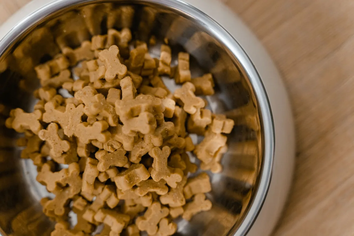
What your dog eats can influence skin integrity, immune function, and the propensity for allergic reactions. While no single “nose‑only” diet exists, choosing a balanced, high‑quality diet supports overall skin health and helps the nose heal faster.
Foods to favor
- Highly digestible protein sources: Chicken, turkey, or salmon provide essential amino acids without excess fat, which can burden the skin’s oil‑producing glands.
- Omega‑3 enriched formulas: Commercial diets labeled “with added fish oil” or “rich in EPA/DHA” have been shown by the AVMA to reduce inflammatory skin conditions.
- Limited‑ingredient or novel‑protein diets: If food allergy is suspected, a diet that eliminates common allergens (beef, dairy, wheat) for 8‑12 weeks can clarify the trigger.
- Balanced vitamins and minerals: Adequate zinc, copper, and vitamin A are critical for epithelial regeneration. Look for AAFCO‑approved foods that list these nutrients in appropriate amounts.
Foods to limit or avoid
- High‑fat treats: Excessive fat can exacerbate skin oiliness and encourage bacterial growth.
- Artificial additives: Preservatives, colorants, and flavor enhancers are common allergens; choose grain‑free or limited‑additive options when possible.
- Table scraps: Human foods often contain spices or ingredients (e.g., onion, garlic) that irritate a dog’s skin.
Prescription therapeutic diets
If your vet identifies a specific underlying disease—such as an autoimmune condition—they may recommend a therapeutic diet designed for immune modulation. Brands like Hill’s Prescription Diet ® c/d or Royal Canin Veterinary Diet ® Dermacomfort are formulated with controlled omega‑3 levels and antioxidants. We do not endorse any particular brand; the choice should be based on your dog’s individual needs and your vet’s guidance.
Feeding tips during recovery
- Offer smaller, more frequent meals to maintain steady energy without overloading the digestive system.
- Warm the food slightly (to body temperature) if your dog’s nose is sensitive to cold.
- Ensure fresh water is always available—hydration aids skin elasticity.
- Transition to a new diet gradually over 7‑10 days to avoid gastrointestinal upset.
In practice, many owners report that adding a modest fish‑oil supplement (as advised by their vet) and switching to a limited‑ingredient kibble reduces nose crusting within two weeks.
Cost and prognosis
Financial considerations are part of every pet‑care decision. Below is a realistic range of what owners typically spend in the United States and United Kingdom for a full work‑up and initial treatment of nasal dermatoses.
| Service | US Estimate | UK Estimate |
|---|---|---|
| Initial exam & basic blood work | $80‑$150 | £60‑£120 |
| Cytology (scrape) & culture | $50‑$100 | £40‑£90 |
| Allergy testing (panel) | $200‑$400 | £150‑£300 |
| Topical prescription medication (2‑week supply) | $30‑$70 | £25‑£60 |
| Systemic immunosuppressant (first month) | $40‑$120 | £35‑£110 |
| Surgical excision (if needed) | $1,200‑$3,500 | £900‑£2,500 |
Prognosis depends on the cause. Allergic or mild infectious dermatoses usually resolve within 2–4 weeks with appropriate therapy. Autoimmune forms can require months of medication but often achieve long‑term control. Nasal tumors carry a more guarded outlook; early detection dramatically improves survival odds.
Prevention and home care
Preventing recurrence is a combination of environmental management, routine grooming, and attentive monitoring.
- Limit sun exposure: For light‑skinned breeds, apply a pet‑safe sunscreen (zinc‑oxide based) before long outdoor sessions.
- Reduce irritants: Keep cleaning products, scented candles, and strong fragrances out of the dog’s nose area. Use fragrance‑free shampoos and wipes.
- Control parasites: Monthly flea and tick preventatives—not just for skin health but also because flea saliva can trigger nasal allergies.
- Regular nose checks: A quick visual once a week—look for dryness, crust, or color changes. This habit catches problems before they flare.
- Maintain humidity: In dry indoor climates, a humidifier can keep nasal mucosa from drying out.
When you notice a mild change, start with gentle cleaning: a damp, soft cloth (no soaps) to remove crust, then apply a thin layer of a vet‑recommended moisturizing ointment. If symptoms persist beyond 48 hours, schedule a veterinary appointment.
Vet’s note
From our vet team: “A nose that looks a little flaky is often just a minor irritation, but when the skin breaks or bleeds, it’s a signal to investigate deeper. Early labs and a simple cytology can differentiate an allergic flare from a bacterial infection, and that distinction drives the treatment plan. Don’t wait for the problem to get worse—early intervention usually means a quicker, less costly recovery.”
Key takeaways
- Dry, cracked, or inflamed noses are common and usually treatable, but sudden swelling or bleeding warrants immediate veterinary care.
- Allergies, infections, autoimmune disease, and trauma are the main drivers of nasal dermatoses; breed and sun exposure influence risk.
- Diagnosis combines visual exam, cytology, cultures, allergy testing, and, when needed, biopsy to rule out tumors.
- Topical anti‑inflammatories, targeted antibiotics/antifungals, and systemic immunosuppressants are the cornerstone treatments.
- Feeding a balanced, omega‑3‑rich diet and avoiding allergens can speed healing and reduce recurrence.
- Regular nose checks, sun protection for light‑skinned breeds, and parasite control are effective prevention strategies.
Myth vs. fact
Myth: A dry nose always means a serious disease.
Fact: Most dry noses are mild irritations; however, persistent cracking or bleeding should be evaluated.
Myth: Nasal dermatitis is contagious to other dogs.
Fact: Most nasal dermatoses are not infectious; bacterial or fungal infections can spread, so good hygiene is still important.
Myth: Home remedies alone can cure an ulcerated nose.
Fact: Severe lesions need veterinary prescription medication and sometimes systemic therapy; home care supports but does not replace professional treatment.
Frequently asked questions
What are the signs of nasal dermatitis in dogs?
Early signs include dryness, flaking, and mild redness; as it progresses you may see cracking, scabbing, bleeding, or ulceration, often accompanied by rubbing or pawing at the nose.
Can a dog’s nose become infected?
Yes—bacterial overgrowth or yeast (Malassezia) can infect the nasal skin, leading to pus‑filled lesions, odor, and discomfort that require targeted antibiotics or antifungal therapy.
How is nasal dermatitis diagnosed by a vet?
The vet starts with a physical exam, then may perform skin cytology, culture, allergy testing, and, if needed, a biopsy to rule out tumors or autoimmune disease.
What treatments are available for dog nose skin issues?
Topical corticosteroids or calcineurin inhibitors, oral antibiotics or antifungals, systemic immunosuppressants for autoimmune cases, and supportive supplements like omega‑3s are common options.
Is nasal dermatitis in dogs a sign of an underlying disease?
It can be; chronic or severe cases often point to allergies, autoimmune disorders, or less commonly, neoplasia, so thorough testing is essential to uncover any hidden conditions.
Can diet affect my dog’s nasal skin health?
Yes—diets rich in omega‑3 fatty acids, high‑quality protein, and balanced vitamins support skin barrier function and can lessen inflammation, especially in allergic or immune‑mediated dermatoses.
Ask the PuppaDogs community
Have a question this article didn’t fully answer? Want to compare notes with other dog owners who’ve been through this? Our community forum is moderated by experienced owners and vets — and answers tend to come fast. Ask in the PuppaDogs community →
References
- American Animal Hospital Association (AAHA) 2022 Canine Skin Health Survey.
- American Veterinary Medical Association (AVMA) Guidelines on Canine Allergic Dermatitis.
- Merck Veterinary Manual, “Dermatitis, Nasal” chapter.
- American College of Veterinary Internal Medicine (ACVIM) Consensus on Immune‑Mediated Skin Disease, 2021.
- World Small Animal Veterinary Association (WSAVA) Nutritional Recommendations for Skin Health, 2023.
- Cornell University College of Veterinary Medicine, “Canine Nasal Tumors” fact sheet.
- UC Davis Veterinary Medicine, “Allergy Testing in Dogs” protocol.
- AAFA (American Academy of Food Allergy) – Canine Food Allergy Overview, 2022.
- Plumb’s Veterinary Drug Handbook – Topical Corticosteroids, 2024 edition.
- Veterinary Dermatology, by J. Scott Weese, 4th edition, 2023.


