





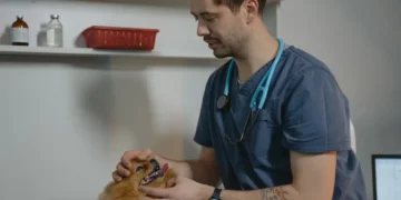
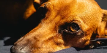

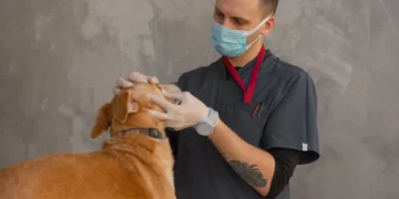






Quick take: Iris cysts are fluid‑filled sacs that develop on the colored part of a dog’s eye. Most are benign and cause only mild irritation, but larger cysts can blur vision or lead to secondary problems. Diagnosis is quick—usually an eye exam with a slit‑lamp or ultrasound—and treatment ranges from observation to laser or surgical removal, often costing a few hundred to a few thousand dollars.
It’s 9 p.m., and you’ve just noticed your normally spry Border Collie squinting at the hallway light. The pup pauses mid‑step, tilts the head, and seems to stare at the ceiling as if something’s “off” in the eye. You glance at the iris and see a small, clear bump that wasn’t there a week ago. Your heart races, and a quick Google search begins. You’re not alone—many owners first spot an iris cyst when a beloved dog’s eye looks a little “off.” Below we walk through exactly what an iris cyst is, how it shows up, when it needs urgent care, and what you can expect from diagnosis, treatment, cost, and everyday management.
Our goal is simple: give you clear, step‑by‑step information so you can feel confident talking with your veterinarian and making the best choices for your dog’s eye health. We’ll cover causes, signs, diagnostic work‑ups, medical and surgical options, nutrition tips, typical costs, and prevention strategies. By the end, you’ll know whether your dog’s cyst is a watch‑and‑wait situation or something that needs prompt intervention.
What is iris cysts?
An iris cyst is a thin‑walled sac filled with clear fluid that forms on the iris—the colored part of the eye that controls how much light enters. In dogs, cysts are usually primary (arising spontaneously) or secondary (linked to trauma, inflammation, or other eye disease. The cyst itself is not cancerous, but because the iris sits right in front of the lens, a large cyst can block vision or cause secondary inflammation.
While exact numbers are scarce, the American College of Veterinary Ophthalmologists (ACVO) notes that iris cysts make up a small fraction of ocular problems seen in practice—roughly 1–2 % of all eye exams in referral centers. They can appear at any age, but senior dogs and breeds with predisposition to eye issues (such as Poodles, Cocker Spaniels, and Bulldogs) are reported more often.
What causes iris cysts?
Most iris cysts are idiopathic, meaning the exact trigger is unknown. However, several risk factors have been identified:
- Genetic predisposition: Certain breeds—especially those prone to uveal (iris) inflammation—show higher rates of cyst formation.
- Eye trauma: A scratch or blunt injury can stimulate fluid‑filled sac development.
- Chronic inflammation: Conditions like uveitis, cataracts, or prior intra‑ocular surgery may lead to secondary cysts.
- Systemic disease: Rarely, hormonal imbalances or neoplastic processes (e.g., ocular melanoma) can mimic cysts.
Because many cases are primary and spontaneous, preventing an iris cyst outright is challenging. Nonetheless, minimizing trauma and promptly treating eye inflammation can reduce secondary cyst risk.
Signs and symptoms
Most owners first notice a subtle change in the eye’s appearance or the dog’s behavior. Early signs often go unnoticed until a closer look is taken.
| Severity | Typical signs |
|---|---|
| Mild | Small, clear bump on iris; slight squinting; occasional “glassy” stare. |
| Moderate | Visible cyst causing pupil distortion; intermittent blinking; mild redness (conjunctival hyperemia). |
| Severe | Large cyst obstructing pupil; persistent redness; tearing; apparent vision loss (bumping into furniture); eye pain (head shaking, pawing at eye). |
In many cases, the cyst itself does not hurt. However, if it enlarges or leads to secondary inflammation, owners may notice:
- Frequent rubbing or pawing at the eye.
- Excessive tearing or discharge.
- Reluctance to play fetch or navigate stairs.
- Changes in eye color or pupil shape.
When to call your vet
Call your regular vet today if you see any of the mild‑to‑moderate signs above—especially squinting, redness, or a visible bump.
Go to an emergency veterinary hospital right now if your dog shows any of these red‑flag symptoms:
- Sudden loss of vision or bumping into objects.
- Severe eye pain (continuous pawing, vocalization, or refusal to open the eye).
- Rapid swelling or a darkening of the iris that could indicate a tumor.
These situations require prompt evaluation to protect vision and rule out more serious conditions like ocular melanoma. This article is for information only and does not replace a hands‑on veterinary exam.
How vets diagnose iris cysts
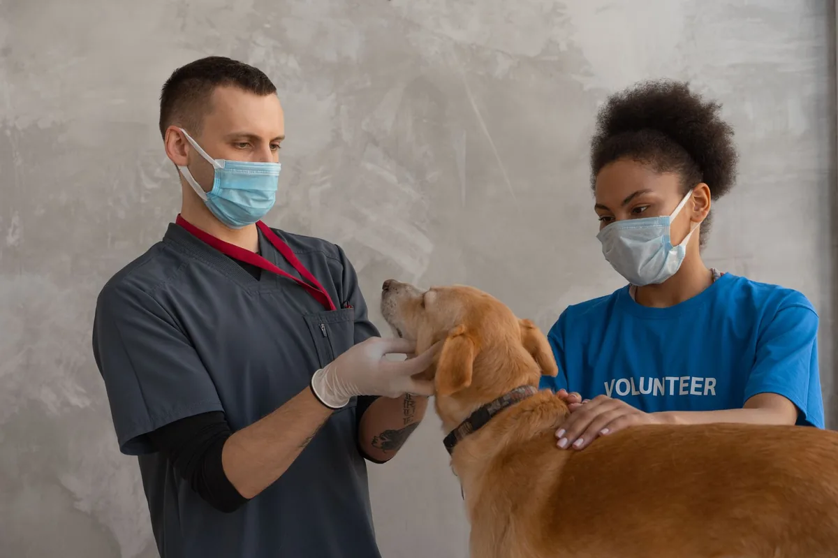
Diagnosing an iris cyst involves a series of non‑invasive steps:
- History and visual exam: Your vet will ask about the onset, any recent injuries, and observe the eye under normal lighting.
- Slit‑lamp biomicroscopy: A specialized microscope provides magnified, illuminated views of the iris, allowing the vet to see the cyst’s size, shape, and internal contents.
- Ultrasound (UBM): High‑frequency ultrasound can assess cyst depth and rule out solid masses.
- Fluorescein staining: A dye helps detect corneal scratches that might accompany a cyst.
- Optional cytology or biopsy: In rare cases where a tumor is suspected, a fine‑needle aspirate may be taken for lab analysis, following guidelines from the AAHA.
These tests are quick, painless, and usually completed in a single appointment. The results guide whether observation, medical therapy, or surgery is needed.
Treatment options
Medical treatment
Most small, asymptomatic cysts are simply monitored. When inflammation or irritation is present, the following medical options are common:
- Topical anti‑inflammatory eye drops: Drugs such as prednisolone acetate or cyclosporine can reduce iris inflammation and may shrink a cyst.
- Systemic anti‑inflammatory medications: Oral NSAIDs (e.g., carprofen) may be prescribed if there’s concurrent ocular discomfort.
- Antibiotic eye drops: Used if secondary bacterial infection is suspected.
These medications are adjuncts; they do not eliminate the cyst but can keep the eye comfortable while you decide on longer‑term plans. Always discuss dosage and duration with your vet.
Supplements and supportive care
While there is no supplement that directly reduces an iris cyst, certain nutrients support overall eye health and may aid recovery after surgery:
- Omega‑3 fatty acids (EPA/DHA): Found in fish‑oil supplements, they help reduce ocular inflammation.
- Antioxidant blends (vitamin C, lutein, zeaxanthin): These nutrients protect retinal cells and are often recommended for dogs with any eye condition.
- Probiotics: Supporting gut health can improve systemic immune response, indirectly helping with inflammatory eye disease.
Ask your vet about adding these to your dog’s diet, especially if surgery is planned.
Surgical procedures
When a cyst threatens vision or grows rapidly, removal is the definitive option. The most common techniques are:
- Laser photocoagulation: A focused laser beam punctures the cyst wall, causing it to collapse. This outpatient procedure often takes under an hour and has a quick recovery.
- Fine‑needle aspiration (FNA): A thin needle draws fluid from the cyst, relieving pressure. It’s less invasive but may require repeat sessions.
- Surgical excision: In rare cases where the cyst is large or atypical, a full‑thickness excision of the iris segment may be performed under general anesthesia.
Post‑operative care typically includes topical antibiotics for a week, anti‑inflammatory drops for 2–3 weeks, and activity restriction (no rough play or jumping) for 7–10 days. Your vet will give a tailored plan based on the chosen technique.
Diet and nutrition
While no specific diet can prevent an iris cyst, feeding a balanced, eye‑supportive diet helps maintain overall ocular health and can aid recovery after surgery. Here are practical guidelines:
| Category | Recommendations |
|---|---|
| Do feed | High‑quality protein sources (chicken, turkey, fish); omega‑3 rich foods (salmon, fish oil); antioxidant‑rich vegetables (carrots, blueberries); AAFCO‑verified complete & balanced kibble. |
| Limit | High‑salt treats, excessive dairy, and foods high in omega‑6 fatty acids (e.g., many commercial treats) that can promote inflammation. |
| Avoid | Raw fish without proper handling (risk of thiamine deficiency), excessive vitamin A supplements, and any diet lacking essential fatty acids. |
For dogs undergoing surgery, a slightly higher caloric intake can support wound healing, but over‑feeding should be avoided to prevent weight‑related eye pressure. Transition any new food gradually over 5–7 days to minimize gastrointestinal upset.
If your dog has a concurrent condition—such as kidney disease, which is common in senior breeds—consider a therapeutic diet formulated for renal support. These diets are lower in phosphorus and protein, easing the workload on the kidneys while still providing adequate nutrition for healing (see AAHA renal diet guidelines).
Our team often recommends adding a fish‑oil supplement (1 % of daily caloric intake) and a canine‑specific multivitamin containing lutein and zeaxanthin for dogs with chronic eye issues. Always discuss supplement choice with your vet to avoid interactions with prescribed medications.
Cost and prognosis
Financial considerations are an important part of any veterinary decision. Below are typical cost ranges for the United States and United Kingdom; actual fees vary by clinic, region, and individual case.
| Service | US estimate | UK estimate |
|---|---|---|
| Initial ophthalmology exam (slit‑lamp, ultrasound) | $150–$250 | £80–£130 |
| Topical anti‑inflammatory prescription (30 days) | $30–$70 | £20–£45 |
| Laser photocoagulation | $500–$1,200 | £300–£700 |
| Surgical excision (including anesthesia) | $1,200–$2,500 | £800–£1,600 |
| Post‑operative meds & rechecks (2 visits) | $100–$250 | £60–£120 |
Prognosis is excellent for most primary cysts—over 90 % retain normal vision after appropriate treatment. Secondary cysts linked to chronic inflammation or tumors have a more guarded outlook, depending on the underlying disease.
Early detection and timely veterinary care are the biggest factors in preserving vision and keeping costs manageable. Many owners find that routine eye checks (once a year for senior dogs) catch cysts before they cause trouble.
Prevention and home care
While you can’t guarantee a cyst won’t form, these habits reduce risk and help you notice problems early:
- Regular eye inspections: During grooming, gently lift the eyelid and look for any bumps, redness, or discharge.
- Protect against trauma: Keep your dog’s play area free of sharp objects; use protective eyewear for dogs that engage in high‑impact activities (e.g., agility).
- Promptly treat eye infections or uveitis: Early anti‑inflammatory therapy can prevent secondary cyst formation.
- Annual veterinary ophthalmology exams: Especially for breeds prone to eye disease, a yearly check can catch subtle changes.
- Maintain a balanced diet with omega‑3s and antioxidants: This supports overall ocular health.
After any procedure, follow your vet’s recheck schedule—usually at 1 week and 4 weeks—to ensure the eye is healing properly. Keep a log of any changes you observe, and bring photos to appointments if possible; visual documentation helps the vet track cyst size over time.
From our vet team: “Most iris cysts are harmless, but they can become a problem if they grow or cause inflammation. The key is early detection—if you notice even a tiny bump or a change in your dog’s eye behavior, schedule a vet visit right away. With modern laser techniques, many cysts are resolved quickly and painlessly, and the outlook for vision is usually very good.”
Key takeaways
- Iris cysts are fluid‑filled sacs on the colored part of the eye; most are benign and cause mild irritation.
- Watch for squinting, redness, tearing, or any visible bump on the iris, and contact your vet promptly.
- Diagnosis is quick using a slit‑lamp exam and ultrasound; treatment ranges from observation to laser or surgical removal.
- Laser photocoagulation is the most common, minimally invasive treatment and often restores normal vision.
- Typical costs in the US run from $150 for an exam up to $2,500 for surgical removal; UK costs are proportionally similar.
- Support eye health with omega‑3s, antioxidants, and regular veterinary eye checks, especially for breeds prone to ocular issues.
Myth vs. fact
Myth: All iris cysts are cancerous and will destroy vision.
Fact: The overwhelming majority of iris cysts are benign; only a small fraction are neoplastic, and most dogs retain normal vision with appropriate care.
Myth: Surgery is always required to fix an iris cyst.
Fact: Small, asymptomatic cysts often just need monitoring, and many respond to laser therapy or anti‑inflammatory drops without invasive surgery.
Myth: You can treat iris cysts at home with over‑the‑counter eye drops.
Fact: Only a veterinarian can safely diagnose an iris cyst and prescribe the correct medication; self‑medicating can worsen inflammation or mask serious disease.
Frequently asked questions
What does an iris cyst look like in a dog’s eye?
An iris cyst appears as a clear or slightly translucent dome‑shaped bump on the iris, often near the pupil. It may be invisible without magnification, but owners sometimes notice a subtle irregularity or a “glassy” stare.
Can iris cysts cause pain or discomfort for dogs?
Most cysts are painless, but if they enlarge or cause inflammation, dogs may squint, rub their eye, or show signs of discomfort. Persistent rubbing or vocalization when the eye is touched signals that veterinary evaluation is needed.
How is an iris cyst diagnosed by a veterinarian?
Vets use a slit‑lamp biomicroscope to view the cyst up close, often supplemented with high‑frequency ultrasound (UBM) to assess depth and rule out solid masses. These tests are quick, non‑invasive, and usually done in a single appointment.
What treatment options are available for iris cysts in dogs?
Treatment ranges from observation for tiny, non‑problematic cysts to topical anti‑inflammatory drops for mild irritation. For larger or vision‑threatening cysts, laser photocoagulation, fine‑needle aspiration, or surgical excision are effective options.
Will my dog lose vision if an iris cyst is left untreated?
If a cyst grows enough to block the pupil or cause secondary inflammation, vision loss can occur. Early detection and treatment dramatically reduce this risk, and most dogs retain full vision after appropriate care.
How much does iris cyst removal surgery typically cost?
In the United States, laser removal usually costs $500–$1,200, while full surgical excision can range from $1,200–$2,500. In the United Kingdom, expect £300–£700 for laser and £800–£1,600 for surgery, plus additional fees for anesthesia and post‑operative meds.
Ask the PuppaDogs community
Have a question this article didn’t fully answer? Want to compare notes with other dog owners who’ve been through this? Our community forum is moderated by experienced owners and vets — and answers tend to come fast. Ask in the PuppaDogs community →
References
- American College of Veterinary Ophthalmologists (ACVO). “Ocular Diseases in Dogs.” Clinical guidelines, 2022.
- American Animal Hospital Association (AAHA). “Guidelines for Ophthalmic Examinations.” 2023.
- Merck Veterinary Manual. “Iris Cysts.” Updated 2023.
- American College of Veterinary Internal Medicine (ACVIM). “Management of Ocular Inflammation.” 2021.
- World Small Animal Veterinary Association (WSAVA). “Nutrition for Ocular Health.” 2022.
- Veterinary Ophthalmology, 5th ed., Gelatt & Gilger, 2020.
- University of California, Davis School of Veterinary Medicine. “Laser Photocoagulation for Canine Iris Cysts.” 2021.
- American Veterinary Medical Association (AVMA). “Pet Owner’s Guide to Eye Care.” 2023.


