
















Quick take: FCE (Fibrocartilaginous Embolism) in dogs is a sudden spinal cord injury caused by a piece of disc material blocking blood flow. It often appears as rapid, non‑painful paralysis that can improve with time, but veterinary care is essential to confirm the diagnosis and start supportive treatment.
It’s 11 p.m., and your usually‑bouncy Border Collie, Max, isn’t answering your call to come outside. He’s lying on the floor, his hind legs limp and unresponsive. You feel his gums—paler than usual—and a wave of panic hits. The internet buzzes with “dog paralysis,” “spinal cord injury,” and “emergency vet.” You wonder if this could be something reversible, or if Max’s life is in danger.
We get it. Sudden loss of mobility is one of the scariest things a dog owner can face. The good news is that FCE is a specific, often‑recognizable cause of acute paralysis, and many dogs make a meaningful recovery with proper care. Below we break down what FCE is, how it differs from other back problems, what you should look for, and how to navigate diagnosis, treatment, cost, and long‑term care.
Read on to learn how to spot the warning signs, what questions to ask your vet, and how to support your dog through the recovery journey. Our dog‑health guide also covers related spinal conditions if you need a broader view.
What is FCE (Fibrocartilaginous Embolism) in dogs?
FCE stands for Fibrocartilaginous Embolism, a non‑traumatic spinal cord injury that occurs when a fragment of fibrocartilage—from the intervertebral disc—breaks off and travels into a spinal blood vessel. The embolus blocks blood flow, causing a localized area of ischemia (lack of oxygen) in the spinal cord. Because the blockage is sudden, the resulting neurological deficit appears within minutes to a few hours.
In dogs, FCE is relatively uncommon but not rare. Studies from veterinary teaching hospitals (e.g., UC Davis, Cornell) estimate that FCE accounts for roughly 5–10 % of acute spinal cord injuries. It can affect dogs of any age, although most cases cluster in adult dogs between 3 and 7 years old.
Unlike intervertebral disc disease (IVDD), which often presents with pain and a gradual decline, FCE typically causes a painless, “drop‑foot” paralysis that is confined to one or two spinal segments. The condition is considered a medical emergency because rapid assessment can influence the chance of recovery.

What causes it?
The exact trigger that makes disc material break loose isn’t always clear, but several risk factors have emerged from clinical observations:
- Age and activity level: Dogs in their prime years that engage in vigorous exercise (jumping, sprinting) are more likely to experience a disc‑related embolus.
- Breed predisposition: Medium‑to‑large breeds such as Border Collies, German Shepherds, Labrador Retrievers, and Doberman Pinschers appear over‑represented in case series.
- Underlying disc degeneration: Even without classic IVDD, age‑related disc changes can weaken the fibrocartilage, making it prone to fragment.
- Sudden trauma or strain: A brief, intense burst of activity—like chasing a ball or climbing stairs—can create the shear force needed for a fragment to dislodge.
- Genetic factors: Some lines may have a hereditary tendency toward disc weakness, though research is still evolving.
Signs and symptoms
Because FCE blocks blood flow at a specific spinal level, the clinical picture is often crisp and localized. The most common pattern is a sudden onset of hind‑limb weakness or paralysis, without obvious pain.
| Severity | Typical Signs |
|---|---|
| Mild | Hind‑leg weakness, stumbling, reduced tail movement, normal gait in front legs. |
| Moderate | Complete hind‑leg paralysis, loss of voluntary urination or defecation, unchanged front‑leg strength, no spinal pain on palpation. |
| Severe | Paralysis of hind legs and possibly one front leg (depending on lesion level), absent reflexes below the lesion, urinary retention, possible respiratory compromise if neck is involved. |
Key distinguishing features from IVDD include:
- No obvious back pain or “hunched” posture.
- Rapid progression (minutes to a few hours) rather than a slow decline.
- Neurologic deficits confined to one spinal segment (often a single “myotome”).
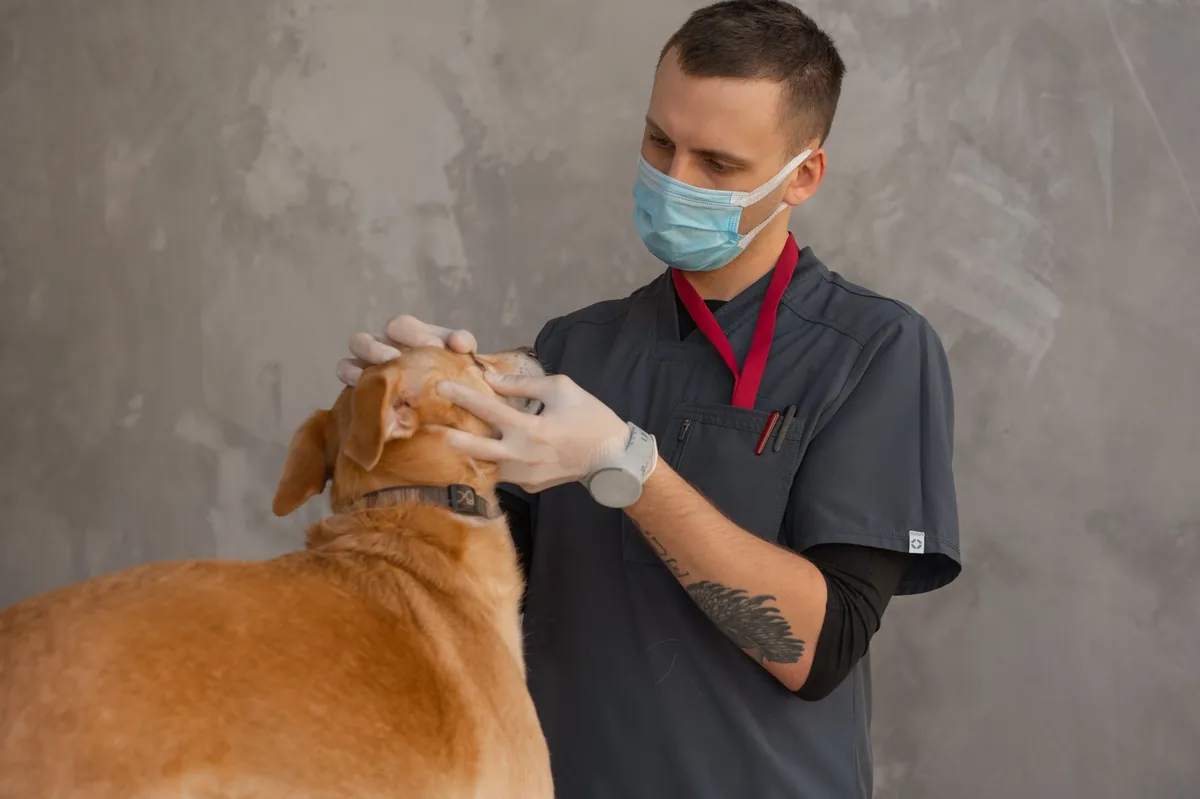
Owners may also notice pale gums, a slightly elevated heart rate, and a lack of response to verbal cues if the lesion affects higher spinal centers.
When to call your vet
Call your regular vet today if:
- Your dog shows sudden hind‑leg weakness or loss of coordination.
- There is any change in bladder or bowel control.
- You notice pale gums, rapid breathing, or a fever.
Go to an emergency veterinary hospital right now if:
- Paralysis has progressed to both hind legs and one front leg within the last hour.
- Your dog cannot urinate at all (no urine output).
- There is a loss of consciousness, severe breathing difficulty, or the neck is involved.
These guidelines are for triage only. Always seek professional veterinary care; this article does not replace a hands‑on exam.
How vets diagnose it
Diagnosing FCE is largely one of exclusion—ruling out other causes of acute paralysis such as IVDD, trauma, or infectious disease. A typical work‑up includes:
- History and neurologic exam: The vet records the exact time of onset, any recent activities, and performs a detailed reflex assessment to localize the lesion.
- Imaging:
- MRI (magnetic resonance imaging): The gold standard for visualizing spinal cord lesions. In FCE, MRI often shows a well‑defined, focal intramedullary hyperintensity without disc protrusion.
- CT scan: Useful if MRI isn’t available, though it may miss subtle cord changes.
- Blood work: CBC and chemistry panels help rule out metabolic or infectious causes and assess overall health before anesthesia.
- Myelography (rare): Contrast dye injected into the spinal canal can highlight compressive disc material, helping differentiate IVDD from FCE.
Because the embolus itself cannot be seen directly, the diagnosis rests on the pattern of sudden, painless paralysis, imaging that shows no disc extrusion, and the exclusion of other diseases.
Treatment options
Medical treatment
Most dogs with FCE are managed medically, especially when the lesion is low‑grade and the dog is stable. Core components include:
- Pain management: NSAIDs (e.g., carprofen) or opioids (e.g., tramadol) are commonly prescribed to keep the dog comfortable.
- Anti‑inflammatory therapy: Corticosteroids such as prednisolone may be used early to reduce spinal cord swelling, though their benefit is debated. Your vet will decide based on the case.
- Neuroprotective agents: Drugs like gabapentin are sometimes added to lessen nerve irritation.
- Physical therapy: Early, gentle passive range‑of‑motion exercises prevent joint contracture and promote circulation. Ask your vet about a tailored rehab plan.
All medication choices should be discussed with your veterinarian; dosage is always weight‑based and tailored to your dog’s condition.
Supplements and supportive care
Evidence supports a few adjuncts that may aid recovery:
- Omega‑3 fatty acids (EPA/DHA): Anti‑inflammatory properties can help reduce secondary spinal cord injury. A high‑quality fish‑oil supplement is often recommended.
- Vitamin B‑complex: B vitamins support nerve metabolism; a balanced B‑complex can be useful during the rehab phase.
- Probiotics: Maintaining gut health is important when a dog is on limited mobility and possibly on antibiotics.
Supplements should never replace prescribed medication, but they can be a helpful part of a comprehensive recovery plan.
Procedures or surgery
Surgery is not the first line for classic FCE because the embolus is lodged within a blood vessel, not a disc that can be removed. However, a few scenarios may call for an operative approach:
- Decompressive surgery: If advanced imaging reveals concurrent disc extrusion or vertebral malformation compressing the cord, a ventral slot or dorsal laminectomy may be performed.
- Spinal stabilization: In cases where the vertebrae are unstable (rare with pure FCE), fixation devices are used.
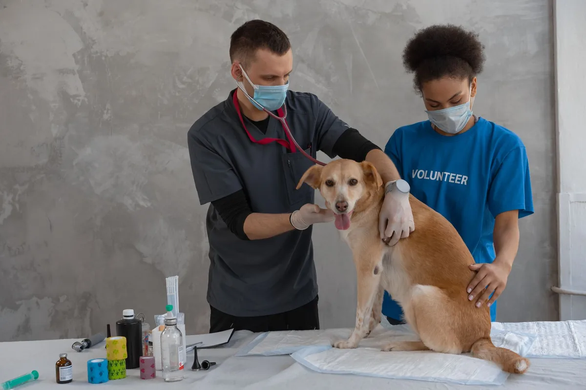
These procedures are typically done at specialty referral hospitals and can cost between $4,000–$8,000 (US) or £3,000–£6,000 (UK), depending on the complexity and length of stay. Recovery after surgery adds an extra few weeks of intensive physiotherapy.
Diet and nutrition
Nutrition plays a supportive role in spinal cord healing. While there is no “FCE‑specific” prescription diet, feeding a balanced, easily digestible diet helps maintain body condition and provides the nutrients needed for nerve repair.
What to feed
- High‑quality protein: Look for meals with named animal proteins (chicken, turkey, salmon) that provide 20–25 % digestible protein. Protein supplies the amino acids essential for nerve regeneration.
- Omega‑3 fatty acids: As mentioned, EPA/DHA reduce inflammation. Many commercial diets now include fish oil; you can also add a supplement.
- Antioxidants: Vitamins E and C, selenium, and beta‑carotene help combat oxidative stress after spinal injury.
- Moderate calories: Over‑feeding can lead to obesity, which slows rehab. Aim for a body condition score of 4–5/9.
What to limit or avoid
- Excessive fat: Very high‑fat meals can cause gastrointestinal upset, especially when the dog is less active.
- Simple carbohydrates: Foods high in corn or wheat may not provide the sustained energy needed for recovery.
- Artificial additives: Preservatives and colorings have no known benefit for nerve healing and can irritate the gut.
Some owners opt for a therapeutic “joint‑support” diet that includes glucosamine, chondroitin, and hyaluronic acid. While these ingredients are primarily for cartilage health, they are harmless and may complement overall recovery.
| Category | Recommended | Limit | Avoid |
|---|---|---|---|
| Protein | High‑quality animal sources (20‑25 % of diet) | — | Low‑quality meat meals |
| Fats | Omega‑3‑rich fish oil | Excess saturated fat | Very high‑fat treats |
| Carbohydrates | Complex grains (sweet potato, brown rice) | Simple carbs (corn, wheat) | — |
| Supplements | EPA/DHA, Vitamin E, B‑complex | — | Unregulated herbal blends |
When transitioning to a new diet, do it gradually over 7–10 days: mix 25 % new food with 75 % old, then increase the new portion every few days. This helps prevent gastrointestinal upset, which can be especially problematic if your dog is already struggling with bladder control.
For specific feeding calculations, our dog calorie calculator can help you determine the right portion size based on weight, activity level, and recovery stage.
Cost and prognosis
Financial considerations are a real part of caring for a dog with FCE. Below is a rough cost breakdown (estimates, may vary by region and clinic):
| Service | US Estimate | UK Estimate |
|---|---|---|
| Initial exam & neurologic work‑up | $150–$300 | £120–£250 |
| MRI of the spine | $1,200–$2,500 | £800–£1,500 |
| Blood panel (CBC, chemistry) | $100–$200 | £80–£150 |
| Hospitalization (3‑day stay) | $500–$1,000 | £400–£800 |
| Physical therapy sessions | $40–$80 per session | £30–£70 per session |
| Optional surgery (if needed) | $4,000–$8,000 | £3,000–£6,000 |
Prognosis varies. Studies from the AAHA and ACVIM indicate that about 60–80 % of dogs regain at least some hind‑leg function with intensive rehab. Small, young dogs and those with lesions limited to one spinal segment tend to recover best. Conversely, dogs with very severe lesions, older age, or delayed treatment may have a poorer outcome.
Recovery timelines are also variable. Most owners see noticeable improvement within 2–4 weeks, but full functional recovery can take 3–6 months of consistent physiotherapy. Patience and regular check‑ins with your vet are crucial.
Prevention and home care
Because FCE is linked to disc health and sudden strain, you can take steps to reduce risk:
- Maintain a healthy weight: Overweight dogs place extra stress on the spine.
- Moderate high‑impact activities: Avoid excessive jumping from heights; use ramps or stairs for older dogs.
- Regular veterinary check‑ups: Early detection of disc degeneration can guide activity modifications.
- Support joint health: Incorporate omega‑3s and joint‑support supplements as part of a balanced diet.
- Safe environment: Keep floors non‑slippery, provide firm bedding, and remove obstacles that could cause sudden twists.
Once your dog is diagnosed, home care focuses on comfort, bladder management, and gentle rehab:
- Bladder care: Use a soft, absorbent pad and monitor urine output. If your dog can’t urinate, a vet‑guided catheter may be needed.
- Passive range‑of‑motion exercises: Gently flex and extend the hind‑leg joints twice daily to prevent stiffness.
- Cold/heat therapy: Apply a cold pack for the first 48 hours if there’s swelling, then switch to warm compresses to improve circulation.
- Hydration and nutrition: Offer water frequently and feed small, frequent meals to aid digestion.
Our expert team can help you design a home‑rehab schedule that matches your dog’s specific lesion level and progress.
From our vet team: FCE can feel like a nightmare, but many dogs bounce back with patience, proper rehab, and a supportive home environment. The key is early veterinary assessment, realistic expectations, and a steady, loving routine that encourages gentle movement without over‑exertion.
Key takeaways
- FCE causes sudden, painless paralysis by blocking spinal blood flow; it’s distinct from disc herniation.
- Immediate veterinary evaluation (especially imaging) is essential to confirm the diagnosis and rule out other emergencies.
- Most dogs are treated medically with pain control, anti‑inflammatories, and structured physiotherapy; surgery is rare.
- Balanced, protein‑rich diets with omega‑3s support nerve healing—avoid excess fats and simple carbs.
- Recovery can take weeks to months; consistent rehab and bladder care dramatically improve outcomes.
- Preventive measures include weight management, moderated activity, and regular vet check‑ups to monitor disc health.
Myth vs. fact
Myth: FCE always requires spinal surgery.
Fact: Most cases are managed medically; surgery is only considered when there’s a concurrent disc extrusion or vertebral instability.
Myth: All dogs with FCE will be permanently paralyzed.
Fact: Up to 80 % regain some hind‑leg function with proper rehab, especially if treated early.
Myth: FCE is contagious or caused by infection.
Fact: It’s a non‑infectious vascular event; there’s no risk of spread to other dogs.
Frequently asked questions
What causes fibrocartilaginous embolism in dogs?
FCE occurs when a piece of fibrocartilage from an intervertebral disc breaks off and blocks a spinal artery, cutting off blood flow to a segment of the spinal cord. The trigger is often a sudden, vigorous movement, especially in dogs with underlying disc degeneration.
Why does my dog suddenly become paralyzed?
Sudden paralysis can result from FCE, intervertebral disc disease, trauma, or spinal infections. FCE is characterized by rapid, painless loss of function limited to one spinal segment, whereas disc disease usually includes back pain.
Is surgery always required for FCE?
No. Because the embolus is lodged in a blood vessel, surgery cannot remove it. Most dogs are treated with pain control, anti‑inflammatory medication, and intensive physiotherapy. Surgery is only considered if imaging shows a co‑existing disc herniation that needs decompression.
How quickly can a dog recover from FCE?
Improvement often begins within 2–4 weeks, but full functional recovery may take 3–6 months of consistent rehab. Younger dogs and those with milder lesions tend to recover faster.
What is the success rate of treatment for FCE?
Current data from the ACVIM suggest that 60–80 % of dogs regain at least partial hind‑leg use with appropriate medical management and physiotherapy. Success depends on lesion severity, age, and how quickly treatment starts.
Are certain dog breeds more at risk for FCE?
Medium‑to‑large breeds such as Border Collies, German Shepherds, Labrador Retrievers, and Doberman Pinschers appear over‑represented in case series. However, any breed can be affected, especially if they have disc degeneration.
Ask the PuppaDogs community
Have a question this article didn’t fully answer? Want to compare notes with other dog owners who’ve been through this? Our community forum is moderated by experienced owners and vets — and answers tend to come fast. Ask in the PuppaDogs community →
References
- AAHA 2023 Canine Neurology Guidelines.
- American College of Veterinary Internal Medicine (ACVIM) Consensus Statement on Acute Spinal Cord Injury, 2022.
- Merck Veterinary Manual, “Fibrocartilaginous Embolism” chapter.
- UC Davis Veterinary Medicine, “Spinal Cord Diseases in Dogs” lecture notes.
- Cornell University College of Veterinary Medicine, “Acute Non‑Traumatic Myelopathies” review.
- World Small Animal Veterinary Association (WSAVA) – Neurology and Rehabilitation resources.
- American Veterinary Medical Association (AVMA) – Guidelines for Pain Management in Dogs.
- Plumb’s Veterinary Drug Handbook – Common analgesics and anti‑inflammatories.
- International Veterinary Rehabilitation Association (IVRA) – Physical therapy protocols for spinal injuries.
- Cost.puppadogs.com – Veterinary service cost estimates (accessed 2026).


