











Quick take: Iris atrophy, also called degeneration of the iris, is a progressive loss of pigment and muscle tone in the eye’s colored ring. It’s most common in older dogs and certain breeds, often painless, but can lead to light sensitivity, glare, and sometimes secondary glaucoma. Diagnosis is quick with a thorough eye exam, and treatment ranges from eye drops to laser therapy; many dogs maintain good vision with proper care.
It’s 9 p.m., and you’ve just noticed your senior Beagle isn’t jumping onto the couch like he used to. When you look closer, the dark ring around his eyes looks faded, almost like a sunset. A quick Google search later, and the term “iris atrophy” pops up, sending a wave of worry through you. You’re not alone—many owners first spot this subtle change while the dog is simply resting.
We understand how unsettling it can be to see a beloved companion’s eyes change. The good news is that iris atrophy is usually painless, and with timely veterinary care most dogs keep a comfortable quality of life. In this article we’ll explain exactly what iris atrophy is, why it happens, how to recognize it, what your vet will do to diagnose it, and the options you have for treatment, cost, and long‑term care.
Read on to learn the signs, the steps you can take tonight, and how to keep your dog’s eyes as bright as ever.
What is degeneration of the iris in dogs?
The iris is the colored part of the eye that controls how much light enters the pupil. In dogs, it contains pigment cells and a thin muscle layer that contracts and relaxes. Degeneration of the iris—also called iris atrophy or iris pigment loss—means these pigment cells and muscle fibers gradually disappear. The result is a lighter‑colored, sometimes misshapen iris that may not respond fully to light changes.
While any dog can develop iris atrophy, it’s most frequently seen in senior dogs over 8 years old. Studies from the American College of Veterinary Ophthalmologists (ACVO) estimate that about 5‑10 % of older dogs show some degree of iris change, though many are mild and go unnoticed.
What causes it?
Iris atrophy can arise from several underlying mechanisms. Below is a concise overview of the main categories.
| Cause | Typical Trigger | Notes |
|---|---|---|
| Age‑related senescence | Natural cellular aging | Most common; gradual pigment loss over years. |
| Genetic predisposition | Breed‑specific genes | Seen in Akita, Siberian Husky, and Shar‑Pei. |
| Trauma or surgery | Eye injury, cataract surgery | Physical damage can accelerate pigment loss. |
| Inflammation (uveitis) | Infectious or immune‑mediated eye inflammation | Chronic inflammation can destroy pigment cells. |
| Systemic diseases | Autoimmune disorders, endocrine issues | Rare, but conditions like hypothyroidism may contribute. |
Signs and symptoms
Because the iris is not innervated with pain fibers, most dogs do not feel discomfort. Instead, owners notice visual or cosmetic changes.
| Severity | Typical Signs |
|---|---|
| Mild | Faded iris color, slight asymmetry, occasional glare in bright light. |
| Moderate | Noticeable light‑sensitivity, pupil may appear larger, occasional “starburst” effect. |
| Severe | Marked iris thinning, irregular pupil shape, secondary glaucoma, reduced vision. |
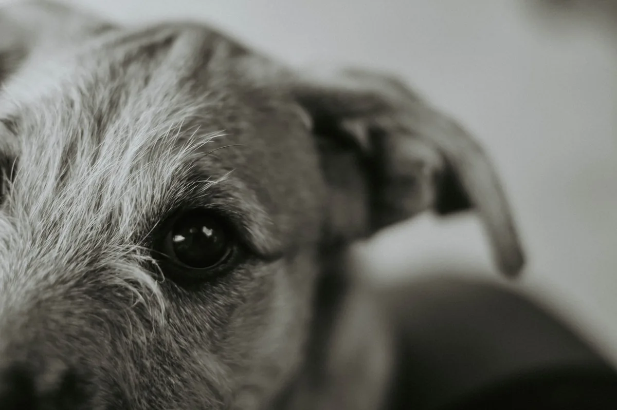
Other clues include a change in eye color when the dog is relaxed versus alert, or a “ghost‑like” appearance of the iris on close inspection. If your dog seems more hesitant to play outside on sunny days, that could be a red flag.
When to call your vet
Call your vet today if you notice:
- Visible fading or uneven coloring of the iris in one or both eyes.
- Increased light sensitivity, especially after a sunny walk.
- Pupil that looks larger than normal or does not constrict fully.
- Any sudden change in vision, such as bumping into objects.
Go to an emergency veterinary hospital right now if you see:
- Redness, swelling, or discharge from the eye.
- Signs of pain – squinting, pawing at the eye, or vocalizing.
- Sudden cloudiness or a deep, painful “glaucoma” episode (eye looks bulging).
These red flags could indicate complications that need immediate attention.
This article is for informational purposes only and does not replace a hands‑on examination by your veterinarian.
How vets diagnose iris atrophy
Diagnosis begins with a detailed history and a full ocular exam. Your vet will:
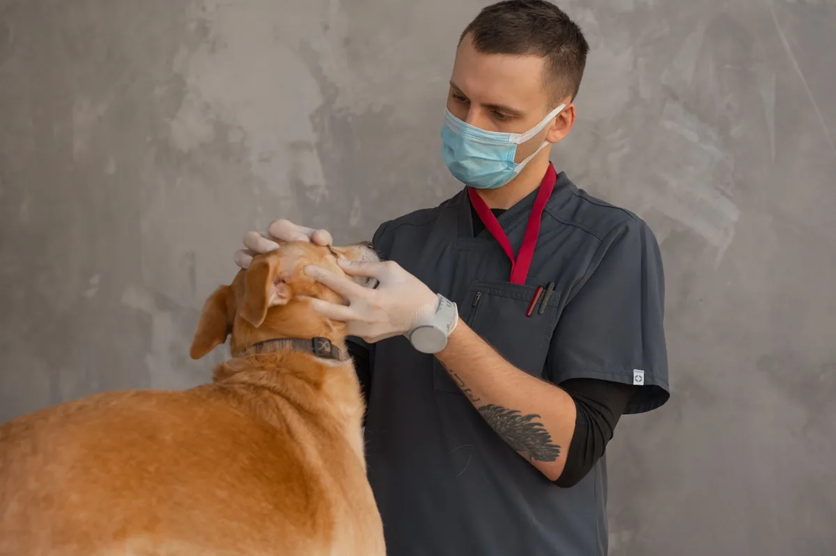
- Perform a slit‑lamp evaluation: a magnified view of the iris, cornea, and anterior chamber to assess pigment loss.
- Measure intraocular pressure (tonometry): to rule out secondary glaucoma, a common complication.
- Use a fluorescein stain: to detect any corneal ulcers that might accompany trauma.
- Take photographs: for baseline comparison over time.
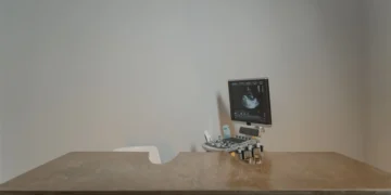
- Consider ocular ultrasound or OCT (optical coherence tomography): if deeper structures need evaluation, especially after cataract surgery.
All of these tests are painless and usually completed in a single visit. The results help the ophthalmologist determine the stage of atrophy and whether any secondary issues are present.
Treatment options
Medical treatment
There is no cure that reverses pigment loss, but several medical approaches can protect vision and reduce discomfort:
- Topical anti‑inflammatory eye drops: such as cyclosporine (Restasis) or corticosteroid formulations to control uveitis if present.
- Pupil‑constricting agents (miotics): like pilocarpine to reduce glare and help maintain a more normal pupil size.
- Glaucoma‑preventing medications: timolol or dorzolamide eye drops if intraocular pressure is trending upward.
- Artificial tears: to keep the ocular surface comfortable, especially in dry environments.
Ask your vet about each option; they’ll tailor the regimen to your dog’s weight, stage of disease, and any concurrent eye conditions.
Supplements and supportive care
While supplements cannot restore lost pigment, they can support overall ocular health:
- Omega‑3 fatty acids (EPA/DHA): found in fish oil, may reduce inflammation and help maintain tear film quality.
- Vitamin C and E: antioxidants that protect ocular cells from oxidative stress, especially in senior dogs.
- Probiotics: promote gut health, which can indirectly influence immune‑mediated eye inflammation.
These are most helpful when the iris atrophy is mild to moderate and the dog has no active infection.
Procedures or surgery
When secondary glaucoma develops or the pupil becomes dangerously dilated, laser or surgical interventions may be recommended:
- Laser iridoplasty: a focused laser creates tiny burns to shrink the iris tissue, improving pupil shape and reducing glare.
- Cyclophotocoagulation: laser treatment that lowers intraocular pressure by partially ablating the ciliary body.
- Glaucoma filtering surgery (tube shunt): reserved for severe pressure spikes that don’t respond to medication.
Procedural costs vary widely; laser iridoplasty typically runs $800‑$1,500 USD (≈ £600‑£1,200), while glaucoma surgery can exceed $2,500 USD (≈ £2,000). Insurance may cover part of the expense, especially if the condition is listed under “eye disease.”
Diet and nutrition
Eye health is closely linked to overall nutrition. While no diet can reverse iris atrophy, feeding a balanced, antioxidant‑rich diet can slow progression and keep the ocular surface comfortable.
Key nutritional principles for dogs with iris atrophy:
- High‑quality protein: Supports the structural integrity of ocular tissues. Look for named meat sources (e.g., chicken, salmon) as the first ingredient.
- Omega‑3 fatty acids: EPA/DHA from fish oil or algae help modulate inflammation. Aim for at least 300 mg EPA + DHA per 10 lb of body weight daily, as recommended by the AAHA.
- Antioxidants: Vitamins C, E, lutein, and zeaxanthin protect retinal cells from oxidative damage. Some therapeutic diets (e.g., renal or ophthalmic formulas) fortify these nutrients.
- Controlled sodium: Reduces the risk of fluid retention that can worsen intraocular pressure.
- Hydration: Adequate water intake keeps the tear film stable; consider a water fountain if your dog prefers running water.
| Food | Do feed | Limit | Avoid |
|---|---|---|---|
| High‑quality kibble or wet food with real meat | Yes – supports protein needs | Low‑fat treats | Highly processed “fillers” (corn, wheat gluten) |
| Fish oil supplement | Yes – omega‑3 source | Excessive doses (>1 g per day) | None if used as directed |
| Vitamin C/E enriched treats | Yes – antioxidant boost | Large quantities (risk of GI upset) | Artificial colorings |
| Raw diet | Possible – ensure balanced | Bone fragments, unbalanced fat | Uncooked pork or fish (risk of parasites) |
When transitioning to a new diet, do it gradually over 7‑10 days to avoid gastrointestinal upset. Mix a small amount of the new food with the current diet, increasing the proportion each day. If your dog is on a prescription ophthalmic diet, follow the feeding guidelines on the label and discuss any adjustments with your vet.
For senior dogs, feeding smaller, more frequent meals can help maintain steady nutrient absorption and keep the eyes comfortable throughout the day.
Cost and prognosis
Financial considerations are an important part of any pet health plan. Below is a rough estimate of typical expenses in the United States and United Kingdom (prices vary by region and clinic).
| Service | US (USD) | UK (GBP) |
|---|---|---|
| Initial ophthalmology consult | $150‑$250 | £120‑£200 |
| Slit‑lamp exam + tonometry | $80‑$130 | £70‑£130 |
| Topical medication (3‑month supply) | $30‑$120 | £25‑£100 |
| Laser iridoplasty | $800‑$1,500 | £600‑£1,200 |
| Glaucoma surgery (tube shunt) | $2,500‑$3,500 | £2,000‑£3,000 |
Most dogs with early‑stage iris atrophy maintain good vision for years, especially when inflammation is controlled. Prognosis is excellent for mild cases and fair to good for moderate cases that respond to treatment. Severe atrophy with secondary glaucoma can lead to vision loss, but timely intervention often preserves enough sight for a comfortable life.
Prevention and home care
While you can’t stop aging, you can reduce risk factors that accelerate iris degeneration:
- Regular eye exams: Annual checks for dogs over 7 years, and biannual for breeds prone to eye disease (e.g., Akita, Siberian Husky).
- Protect from trauma: Keep your dog away from rough play that could injure the eye, and use protective eyewear for dogs that work in dusty environments.
- Control inflammation: Promptly treat any uveitis, conjunctivitis, or corneal ulcers; chronic inflammation speeds pigment loss.
- Maintain a healthy weight: Obesity can increase intraocular pressure and inflammation.
- Use flea/tick preventatives: Some vector‑borne diseases (e.g., Bartonella) can cause eye inflammation.
At home, you can monitor your dog’s eyes by gently lifting the eyelid and checking for changes in color or pupil shape. A quick “gently press the corner of the eye with a clean fingertip” test can reveal whether the pupil constricts normally. If you notice any sudden change, contact your vet right away.
From our vet team: “We often see owners panic over a faded iris, but most cases are painless and manageable. The key is early detection, consistent medication, and protecting the eye from secondary glaucoma. If you keep a regular eye‑health schedule and ask about anti‑inflammatory drops at the first sign of change, your senior dog can enjoy clear vision for many more happy years.”
Key takeaways
- Iris atrophy is a common, usually painless condition in older dogs that causes the iris to lose pigment and muscle tone.
- Watch for faded iris color, increased light sensitivity, or an unusually large pupil; these are early warning signs.
- Prompt veterinary evaluation—including slit‑lamp exam and pressure testing—helps prevent complications like glaucoma.
- Medical management (anti‑inflammatory drops, miotics, glaucoma meds) and, when needed, laser therapy can preserve vision.
- Feed a balanced diet rich in high‑quality protein, omega‑3s, and antioxidants to support overall eye health.
- Regular eye check‑ups, trauma avoidance, and controlling inflammation are the best ways to slow progression.
Myth vs. fact
Myth: Iris atrophy always leads to blindness.
Fact: Most dogs retain functional vision; blindness only occurs if secondary glaucoma develops or the atrophy is extremely severe.
Myth: There’s a quick “cure” for faded irises.
Fact: Treatment focuses on managing symptoms and preventing complications; pigment loss itself cannot be reversed.
Myth: Only purebred dogs get iris atrophy.
Fact: While certain breeds have higher risk, mixed‑breed and rescue dogs can develop the condition, especially as they age.
Frequently asked questions
What are the early signs of iris atrophy in dogs?
The first clue is usually a subtle fading of the iris color, often noticed when the dog is relaxed. Light sensitivity and a slightly larger pupil may follow. Owners often spot these changes during a routine grooming or by comparing photos over months.
Can iris degeneration be reversed in dogs?
Currently, there is no way to restore lost pigment. However, anti‑inflammatory eye drops and laser iridoplasty can improve pupil function and reduce glare, effectively “reversing” the functional impact for many dogs.
Is iris atrophy hereditary in certain breeds?
Yes. Breeds such as Akita, Siberian Husky, and Shar‑Pei have a higher genetic predisposition. If you own one of these breeds, schedule annual ophthalmology exams starting around age six.
How does iris atrophy affect a dog’s vision?
The iris controls light entry; when it thins, the pupil may not constrict properly, leading to glare and reduced contrast. Most dogs adapt, but severe cases can cause difficulty seeing in bright light or navigating stairs.
What tests does a vet use to confirm iris atrophy?
Veterinarians perform a slit‑lamp exam, measure intraocular pressure with a tonometer, and may use fluorescein staining to rule out corneal ulcers. Photographic documentation helps track progression over time.
What is the typical cost of surgery or medication for iris atrophy?
Medication (eye drops) for three months usually costs $30‑$120 USD. Laser iridoplasty ranges $800‑$1,500 USD, while glaucoma surgery can exceed $2,500 USD. Insurance may cover part of these expenses if the condition is listed under “eye disease.”
Ask the PuppaDogs community
Have a question this article didn’t fully answer? Want to compare notes with other dog owners who’ve been through this? Our community forum is moderated by experienced owners and vets — and answers tend to come fast. Ask in the PuppaDogs community →
References
- American College of Veterinary Ophthalmologists (ACVO). “Iris Atrophy in Dogs” clinical guidelines, 2022.
- American Animal Hospital Association (AAHA). “Nutritional Management of Ocular Health,” 2023.
- Merck Veterinary Manual. “Eye Diseases – Iris Disorders” chapter, 2021.
- American Veterinary Medical Association (AVMA). “Glaucoma in Dogs” fact sheet, 2022.
- Cornell University College of Veterinary Medicine. “Senior Dog Eye Health” client handout, 2023.
- World Small Animal Veterinary Association (WSAVA). “Guidelines for Ophthalmic Examinations,” 2022.
- American College of Veterinary Internal Medicine (ACVIM). “Management of Uveitis” consensus statement, 2021.
- Veterinary Ophthalmology textbook, Gelatt & Gilger, 6th edition, 2020.


