


Quick take: Brain tissue underdevelopment in dogs is a rare, usually congenital condition where the brain never reaches its normal size or complexity. It can cause lifelong neurologic signs, but many dogs lead happy, manageable lives with proper care, therapy, and nutrition.
It’s 11 p.m. and your 3‑month‑old Boxer puppy is still sleeping in the corner, eyes half‑closed, and you notice he doesn’t respond to his name the way his littermates do. You glance at his tiny, floppy ears and wonder if something deeper is wrong. You’re not alone—many owners first spot subtle neurological quirks that turn out to be brain tissue underdevelopment, also called cerebral hypoplasia.
We understand the flood of questions that follow: “Is this life‑threatening?” “Can anything be done?” “Will I need to spend a fortune?” Below we break down what brain tissue underdevelopment is, why it happens, how it shows up, and what you can realistically expect in terms of diagnosis, treatment, cost, and everyday care.
By the end of this guide you’ll know the red‑flag signs that merit an immediate veterinary call, the diagnostic steps your vet will likely take, and practical steps you can take at home to give your dog the best quality of life.
What is brain tissue underdevelopment in dogs?
Brain tissue underdevelopment—sometimes called cerebral hypoplasia—is when the brain’s structures (cortex, cerebellum, or brainstem) never fully form during fetal or early post‑natal growth. Unlike brain atrophy, which is a loss of previously normal tissue (often due to aging, injury, or disease), hypoplasia is a developmental shortfall that is present from birth.
It’s relatively uncommon, affecting an estimated 1–2 % of puppies with known congenital anomalies, according to the American College of Veterinary Internal Medicine (ACVIM). The condition can be isolated (affecting only the brain) or part of a broader syndrome that includes eye, skeletal, or cardiac defects.
What causes brain tissue underdevelopment?
The underlying causes fall into three broad categories: genetic, environmental, and infectious/toxic. Below is a concise overview.
| Category | Typical Causes |
|---|---|
| Genetic | Inherited mutations (e.g., LHX1, PAX6) seen in certain breeds; chromosomal abnormalities; inbreeding |
| Environmental/Nutritional | Maternal malnutrition (especially folic acid, iodine, omega‑3 fatty acids); exposure to teratogenic drugs (e.g., phenobarbital, certain antibiotics) during gestation |
| Infectious/Toxic | Canine herpesvirus, parvovirus, or toxoplasmosis in utero; maternal exposure to lead, mycotoxins, or certain pesticides |
Breed predisposition is modest but notable. Small breeds such as Chihuahuas, Pomeranians, and Miniature Schnauzers have been reported more often in case series, likely because breeders sometimes emphasize conformation over health. Mixed‑breed puppies can be affected as well.
Signs and symptoms
Because the brain never fully matures, the clinical picture can range from subtle to severe. Early signs often masquerade as “just being a puppy.” Recognizing the pattern helps you intervene sooner.
| Severity | Typical Signs |
|---|---|
| Mild | Delayed walking or stumbling, poor coordination (ataxia), slow response to commands, occasional head tilts |
| Moderate | Persistent seizures or focal twitching, pronounced tremors, difficulty swallowing (dysphagia), vision problems, abnormal eye movements (nystagmus) |
| Severe | Progressive paralysis, severe seizures, inability to stand, chronic pain, failure to thrive, respiratory compromise |
Behavioral red flags include a lack of curiosity, failure to learn basic cues, and an unusually “spaced‑out” stare. Some owners notice that their puppy prefers to stay in one spot, avoids stairs, or seems overly clumsy during play.
When to call your vet
Call your vet today if you notice:
- Persistent stumbling, unsteady gait, or repeated head tilts.
- Any seizure activity, even a single event.
- Difficulty eating, swallowing, or drinking.
- Abnormal eye movements or vision loss.
Go to an emergency veterinary hospital right now if you see:
- Sudden collapse or inability to stand.
- Continuous seizures lasting more than 5 minutes (status epilepticus).
- Severe respiratory distress or choking.
These recommendations are for information only and do not replace professional veterinary care.
How vets diagnose brain tissue underdevelopment
Diagnosis begins with a thorough history—maternal health, exposures, litter information—and a detailed neurological exam. The vet will assess gait, reflexes, cranial nerve function, and behavior.
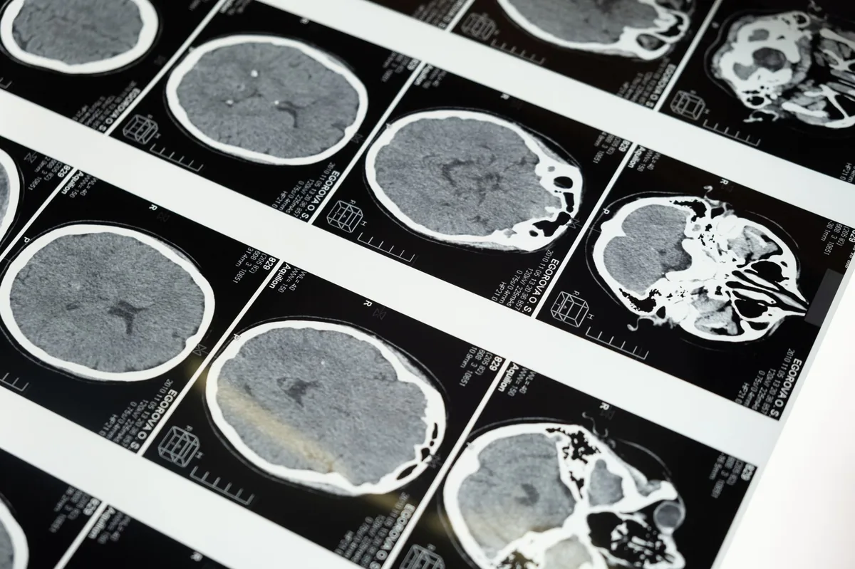
Imaging is the cornerstone:
- MRI (Magnetic Resonance Imaging): Provides high‑resolution images of brain architecture, revealing reduced brain volume, enlarged ventricles, or absent structures. MRI is the gold standard for confirming hypoplasia.
- CT (Computed Tomography): Useful when MRI isn’t available; can show structural deficits but with less soft‑tissue detail.
Additional tests may include:
- Genetic panels (when a hereditary breed risk is suspected).
- Blood work to rule out metabolic causes of seizures or ataxia.
- CSF (cerebrospinal fluid) analysis if infection is a concern.
All findings are interpreted together to differentiate true underdevelopment from acquired brain atrophy or other neurologic disorders.
Treatment options
Medical treatment
There is no cure that can make the brain grow to normal size, but supportive medicines can manage symptoms:
- Anticonvulsants: Drugs like phenobarbital or levetiracetam are commonly prescribed to control seizures. Ask your vet about this option.
- Anti‑inflammatory agents: Corticosteroids (e.g., prednisone) may reduce cerebral edema in acute phases. Ask your vet about this option.
- Neuroprotective supplements: Omega‑3 fatty acids (EPA/DHA) and antioxidants such as vitamin E can support neuronal health. Ask your vet about this option.
Supplements and supportive care
Evidence supports a few adjuncts that can improve quality of life:
- Omega‑3 fatty acids: 1000 mg of combined EPA/DHA per 10 lb of body weight daily has been shown to reduce seizure frequency in some dogs (Cornell Veterinary Medicine). Ask your vet about this option.
- Probiotics: Strains like Enterococcus faecium help maintain gut health, which can indirectly lessen neurologic stress.
- Vitamin B complex: Supports metabolic pathways critical for neural function; especially useful if a nutritional deficiency contributed to the condition.
Procedures or surgery
In rare cases where a specific structural defect (e.g., a persistent fontanelle causing cerebrospinal fluid leakage) is identified, corrective surgery may be recommended. Recovery typically involves 2–4 weeks of restricted activity, and costs can range from $2,500–$5,000 in the United States.
Diet and nutrition
While diet cannot reverse an existing structural deficit, a well‑balanced, brain‑supportive nutrition plan can minimize secondary problems and help your dog thrive.
| Do feed | Limit | Avoid |
|---|---|---|
| High‑quality protein (chicken, turkey, fish) with digestible amino acids | Excessive grain fillers (>30 % of diet) | Artificial preservatives, colors, and flavors |
| Omega‑3‑rich foods (salmon, sardines, fish oil supplements) | High‑fat treats (>15 % of daily calories) | Excessive sodium and processed meats |
| Complex carbohydrates (sweet potato, pumpkin) for steady energy | Very high‑calorie “weight‑gain” formulas unless needed | Simple sugars (white rice, corn syrup) |
Key nutritional considerations:
- Highly digestible protein: Keeps muscle mass intact, which is crucial for dogs that may have motor deficits.
- Omega‑3 fatty acids: EPA and DHA are integral to neuronal membrane fluidity; they also have anti‑inflammatory properties.
- Antioxidants: Vitamin E, selenium, and beta‑carotene can mitigate oxidative stress that may exacerbate neurologic decline.
- Balanced micronutrients: Adequate levels of B‑vitamins (especially B12 and folate) support myelin formation and overall brain metabolism.
If your dog has seizures, a prescription diet formulated for neurological health—such as a “neurology” formula approved by the AAHA—may be beneficial. Discuss options with your vet; they can help transition slowly over 7–10 days to avoid gastrointestinal upset.
Cost and prognosis
Financial planning is a realistic concern for many families. Below are typical cost ranges in the United States and United Kingdom (prices vary by clinic and region).
| Service | US (USD) | UK (GBP) |
|---|---|---|
| Initial neurology consult | $150–$300 | £100–£200 |
| MRI scan (brain only) | $1,200–$2,500 | £800–£1,500 |
| Genetic testing panel | $300–$600 | £250–£500 |
| Anticonvulsant medication (first year) | $200–$500 | £150–£350 |
| Specialty diet (annual) | $400–$800 | £300–£600 |
| Surgery (if indicated) | $2,500–$5,000 | £1,800–£3,500 |
Prognosis depends on severity. Dogs with mild hypoplasia and well‑controlled seizures can live a normal lifespan, often 12–15 years. Severe cases with progressive neurologic decline may have a reduced life expectancy of 2–4 years, though quality of life can still be high with proper management.
Prevention and home care
Because many cases are congenital, primary prevention focuses on maternal health and responsible breeding practices.
- Breeding considerations: Choose studs and dams with health clearances, avoid inbreeding, and request genetic screening where available (e.g., via our experts).
- Maternal nutrition: Feed pregnant bitches a balanced diet rich in folic acid, iodine, and omega‑3s; consider a prenatal supplement under veterinary guidance.
- Toxin avoidance: Keep pregnant dogs away from pesticides, lead‑containing paints, and certain medications known to be teratogenic.
- Early detection: Monitor puppies for developmental milestones (walking, eye tracking, response to sound) and schedule a neurology exam if delays are noted.
At home, maintain a safe environment: non‑slippery flooring, low‑step entryways, and supervised play areas reduce the risk of injury for dogs with coordination problems.
From our vet team: “If your puppy shows even a subtle gait abnormality, we recommend a neurologic exam sooner rather than later. Early imaging can clarify whether we’re dealing with hypoplasia or another treatable issue, and that knowledge guides the most effective supportive plan.”
Key takeaways
- Brain tissue underdevelopment is a congenital condition where the brain never reaches normal size; it differs from brain atrophy, which is loss of previously normal tissue.
- Early signs include delayed walking, ataxia, seizures, and abnormal eye movements—any of these merit a veterinary call.
- Diagnosis relies on MRI (the gold standard), neurological exams, and sometimes genetic testing; CT is a secondary option.
- Management focuses on seizure control, nutritional support (especially omega‑3s), and safe home environments; surgery is rare but may help specific structural defects.
- Costs can add up—expect $1,500–$3,000 for initial work‑up and ongoing medication and diet expenses; budgeting early helps avoid surprises.
- Preventing the condition centers on responsible breeding, proper maternal nutrition, and avoiding teratogenic exposures during pregnancy.
Myth vs. fact
Myth: Brain tissue underdevelopment always leads to a short, painful life.
Fact: Many dogs with mild hypoplasia live full, happy lives with seizure control and supportive care; prognosis varies widely.
Myth: MRI is a luxury test and not necessary for diagnosing brain issues.
Fact: MRI provides the most accurate view of brain structure and is essential for confirming hypoplasia versus other neurologic disorders.
Myth: Once diagnosed, the condition cannot be managed at home.
Fact: Consistent medication, diet, and a safe environment can dramatically improve daily function and quality of life.
Frequently asked questions
What are the early signs of brain tissue underdevelopment in a dog?
Early signs often include delayed walking, unsteady gait, head tilts, and reduced responsiveness to commands. Seizure activity—whether a single episode or recurring—should also trigger a vet visit.
Can brain tissue underdevelopment be diagnosed with an MRI?
Yes. MRI is the gold‑standard imaging method for visualizing reduced brain volume and structural anomalies associated with hypoplasia.
Is brain tissue underdevelopment hereditary in certain breeds?
Some breeds show a higher incidence, suggesting a genetic component. While no single breed is guaranteed to develop the condition, breeds like Chihuahuas, Pomeranians, and Miniature Schnauzers have been reported more often in case series.
What treatment options are available for dogs with underdeveloped brain tissue?
Treatment focuses on symptom management: anticonvulsants for seizures, anti‑inflammatory drugs if edema is present, omega‑3 supplements, and, in rare cases, corrective surgery for specific structural defects.
How long can a dog live with brain tissue underdevelopment?
Life expectancy ranges from a few years in severe cases to a normal lifespan (12‑15 years) in milder forms, especially when seizures are well controlled and supportive care is provided.
Will my dog need lifelong medication for brain tissue underdevelopment?
Many dogs require ongoing anticonvulsant therapy to manage seizures. The need for lifelong medication depends on severity; some dogs may taper off under veterinary supervision if seizure control is achieved.
Ask the PuppaDogs community
Have a question this article didn’t fully answer? Want to compare notes with other dog owners who’ve been through this? Our community forum is moderated by experienced owners and vets — and answers tend to come fast. Ask in the PuppaDogs community →
References
- American College of Veterinary Internal Medicine (ACVIM). “Cerebral Hypoplasia in Dogs.” 2022 guideline.
- American Animal Hospital Association (AAHA). “Neurology Standards for Companion Animals.” 2023 edition.
- Merck Veterinary Manual. “Brain Development Disorders.” Updated 2024.
- Cornell University College of Veterinary Medicine. “Omega‑3 Fatty Acids in Canine Neurology.” 2021 research review.
- World Small Animal Veterinary Association (WSAVA). “Guidelines for Genetic Testing in Companion Animals.” 2022.
- Veterinary Information Network (VIN). “Diagnostic Imaging of Canine Cerebral Hypoplasia.” 2023.
- American Veterinary Medical Association (AVMA). “Seizure Management in Dogs.” 2023 policy.
- AKC Canine Health Foundation. “Breed‑Specific Neurologic Disorders.” 2022 report.
