


Quick take: Bleeding of the retina in the eye in dogs is a sign that something inside the eye is leaking blood, often due to trauma, high blood pressure, or clotting problems. If you notice sudden vision changes, dark spots, or a red‑tinged pupil, contact your veterinarian promptly—early diagnosis can improve the chance of preserving sight.
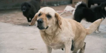
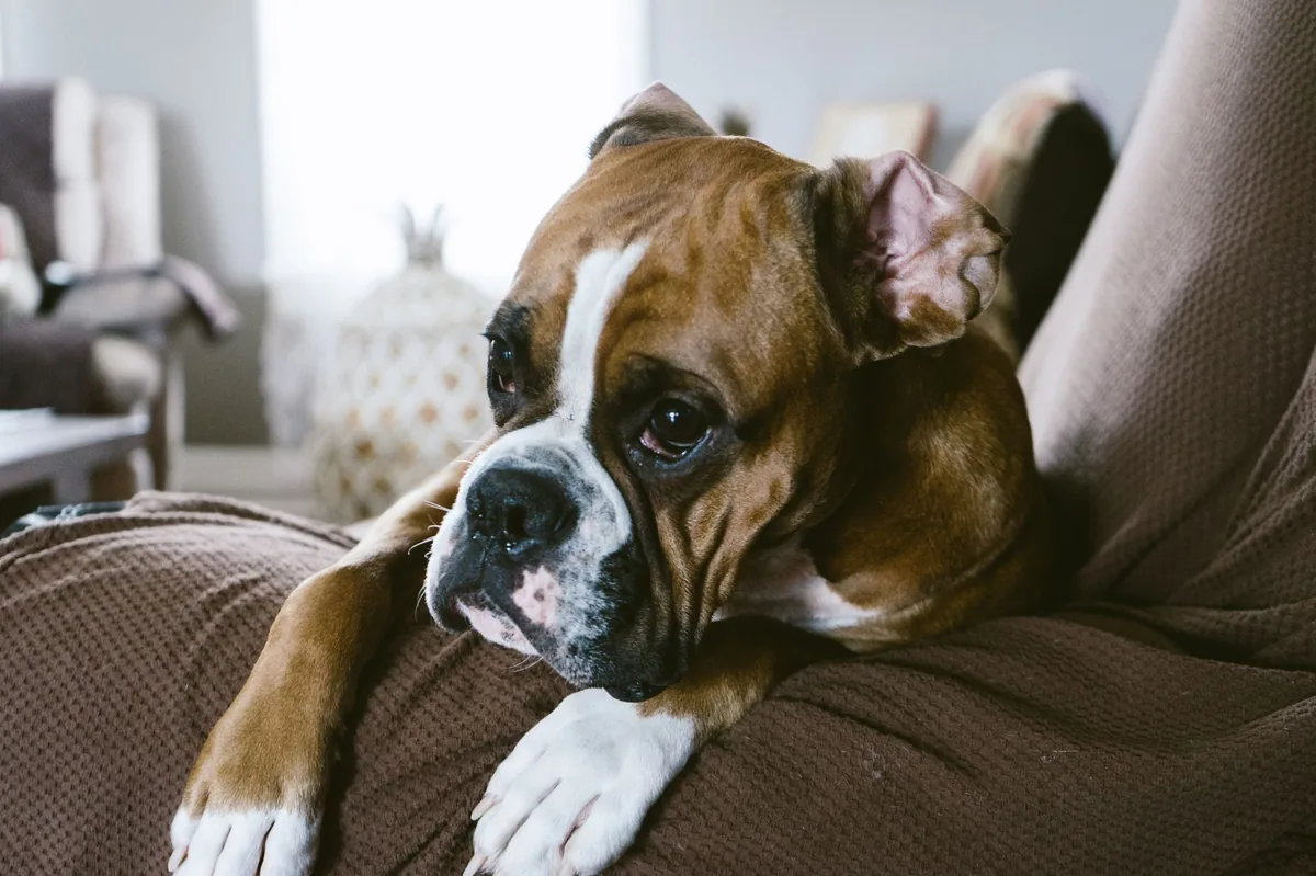
It’s 11 p.m., and you’re sitting on the couch when your usually bright‑eyed Boxer doesn’t chase the ball like usual. Instead, he pauses, his head tilted, and you notice a faint reddish halo around his pupil. Your heart races, and a quick Google search later you’re reading about “bleeding of the retina in the eye in dogs.” You wonder: Is this an emergency? Will he go blind? Can you do anything at home?
We get it—seeing blood where it shouldn’t be is scary. The good news is that retinal hemorrhage is often treatable, especially when caught early. In this guide we’ll explain what a retinal bleed is, why it happens, how vets find it, and what you can expect from medical or surgical care. We’ll also cover diet tweaks, cost estimates, and everyday steps to keep your dog’s eyes healthy.
What is bleeding of the retina in the eye in dogs?
The retina is the thin, light‑sensitive layer at the back of the eye that turns visual images into nerve signals. When blood vessels inside this layer rupture, blood pools in the retinal tissue—a condition called retinal hemorrhage. The bleed can appear as a small dot, a larger patch, or a diffuse red tint that obscures vision.
Retinal bleeding is relatively uncommon but not rare. Studies from the American College of Veterinary Ophthalmologists (ACVO) suggest that 1–3 % of dogs examined for eye problems have some form of retinal hemorrhage. Small, isolated bleeds often resolve on their own, while larger or repeated bleeds may signal a serious underlying disease.
What causes bleeding of the retina in dogs?
Several systemic and local factors can weaken retinal vessels enough to leak. The most frequent culprits include:
| Cause / Risk factor | How it leads to a retinal bleed |
|---|---|
| Trauma | Blunt or penetrating eye injury tears tiny vessels. |
| Systemic hypertension (high blood pressure) | Elevated pressure stresses vessel walls, especially in older dogs. |
| Coagulopathies (bleeding disorders) | Inherited or acquired clotting defects prevent normal clot formation. |
| Thrombocytopenia (low platelets) | Fewer platelets mean bleeding stops more slowly. |
| Infectious diseases (e.g., ehrlichiosis, heartworm) | Inflammation and vessel damage from parasites or bacteria. |
| Neoplasia (eye tumors) | Tumor growth can disrupt normal vasculature. |
Breeds such as the Boxer, Doberman Pinscher , and Miniature Schnauzer are reported more often in studies of hypertension‑related retinal bleeds, but any dog can be affected.
Signs and symptoms
Because the retina is part of the visual system, the first clues are usually changes in how your dog sees or behaves. Early signs are subtle; later signs suggest more extensive bleeding.
| Severity | Typical signs |
|---|---|
| Mild | Brief stare, slight head tilt, occasional bumping into furniture. |
| Moderate | Dark spots or “floaters” in vision, reduced interest in play, hesitation on stairs. |
| Severe | Sudden blindness, noticeable red or cloudy pupil, blood‑tinged discharge, persistent eye pain. |
Other observable cues include:
- Pale or red‑tinged gums (a sign that the bleed may be part of a systemic issue).
- Excessive tearing or ocular discharge.
- Changes in pupil size—often a smaller, irregular pupil on the affected side.
When to call your vet
Call your vet today if you notice any of the following:
- Sudden head tilt or loss of coordination.
- Visible red spots or a reddish tint in the pupil.
- Persistent eye discharge or tearing.
- Changes in appetite or energy that accompany the eye signs.
Go to an emergency veterinary hospital right now if you see:
- Sudden, complete blindness in one or both eyes.
- Severe eye pain—your dog pawing at the eye, vocalizing, or flinching.
- Bleeding that looks like fresh blood leaking from the eye surface.
This article is for informational purposes only and does not replace professional veterinary care.
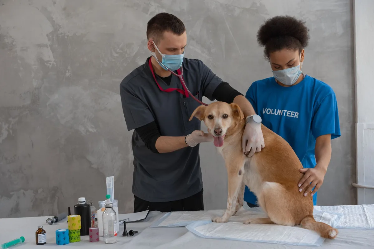
How vets diagnose retinal bleeding in dogs
Diagnosis begins with a thorough history: recent injuries, known blood‑pressure issues, or medications that affect clotting. The physical exam includes a careful eye check using a slit‑lamp microscope and an ophthalmoscope.
Key diagnostic tools:
- Fundic (retinal) photography—captures images of the back of the eye so the vet can see the exact size and location of the bleed.
- Ultrasound (B‑scan)—helps evaluate the eye’s internal structures when cataracts or other opacity block direct view.
- Complete blood count (CBC) and coagulation panel—detects anemia, low platelets, or clotting factor deficiencies.
- Blood pressure measurement—identifies hypertension, a common driver of retinal bleeds in older dogs.
- Abdominal ultrasound or thoracic imaging—looks for systemic disease (e.g., kidney disease, heart disease) that may be causing high blood pressure.
All of these tests together let the veterinarian pinpoint the cause and decide whether medical management or surgery is appropriate.
Treatment options
Medical treatment
When the bleed is small and the underlying cause can be controlled, most vets start with medication. Common drug classes include:
- Antihypertensives such as amlodipine or enalapril—help lower systemic blood pressure to prevent further vessel rupture.
- Anti‑inflammatory agents like prednisolone or non‑steroidal anti‑inflammatories (NSAIDs) to reduce swelling around the retina.
- Anticoagulant reversal agents (e.g., vitamin K1) if a clotting defect from rodenticide exposure is identified.
- Antibiotics or antiparasitics when an infectious disease (e.g., ehrlichiosis) is the trigger.
Ask your vet about these options; dosages are always weight‑based and tailored to your dog’s specific situation.
Supplements and supportive care
While supplements cannot replace proper medical treatment, a few have shown benefit in supporting ocular health:
- Omega‑3 fatty acids (EPA/DHA)—help reduce inflammation and may improve retinal microcirculation.
- Antioxidant blends (vitamin C, vitamin E, lutein)—support retinal cell health, especially after a bleed.
- Probiotics—maintain gut health, which can indirectly influence systemic inflammation.
These should be discussed with your vet, as excessive amounts can interfere with clotting or other medications.
Surgical and procedural options
Large or persistent bleeds, especially those that threaten vision, may require surgery. The most common procedure is a pars plana vitrectomy (PPV), where the vet removes the vitreous gel and any blood‑filled membranes to restore a clear visual path.
What to expect:
- Procedure—performed under general anesthesia, lasting 1–2 hours.
- Recovery—dogs usually stay in the hospital 24 hours for monitoring, then go home with eye drops and a protective cone.
- Cost—in the United States, PPV typically ranges from $2,500 to $4,500; in the United Kingdom, expect £1,800 to £3,200.
Diet and nutrition
Nutrition plays a subtle but important role in supporting eye health and managing the systemic conditions that often accompany retinal hemorrhage. While no single “retinal‑bleed diet” exists, feeding a balanced, antioxidant‑rich diet can help protect retinal vessels.
Foods to favor
- High‑quality protein sources (chicken, turkey, fish) that provide essential amino acids for tissue repair.
- Omega‑3‑rich fish oils or flaxseed—support anti‑inflammatory pathways.
- Colorful vegetables (carrots, pumpkin, spinach) that supply beta‑carotene and lutein, both known to support retinal cells.
- Low‑sodium kibble or wet food—helps keep blood pressure in check, especially for senior dogs prone to hypertension.
Foods to limit or avoid
- High‑salt treats or table scraps that can raise blood pressure.
- Excessive butter or fatty cuts that may worsen inflammation.
- Artificial preservatives and dyes, which have been linked to ocular irritation in some studies.
For dogs with an underlying kidney or heart disease, a therapeutic diet (often labeled “renal” or “cardiac”) may be recommended. These diets are formulated to reduce sodium, phosphorus, and protein load while still delivering essential nutrients.
When transitioning to a new diet, do it gradually over 7–10 days: mix 25 % new food with 75 % old food, then increase the new portion every few days. This helps maintain gut health and reduces the chance of gastrointestinal upset.
| Dietary component | Do feed | Limit | Avoid |
|---|---|---|---|
| Protein | High‑quality animal protein | Excessive raw meat diets without balance | Low‑quality filler proteins |
| Fat | Omega‑3 sources | Large amounts of saturated fat | Trans fats |
| Carbohydrates | Whole grains or sweet potatoes | Highly processed carbs | Simple sugars |
| Salt | Moderate (≤0.3 % of diet) | High‑sodium treats | Table scraps with added salt |
Remember, any diet change should be discussed with your veterinarian, especially if your dog is on medication that may interact with certain nutrients (e.g., fish oil and anticoagulants).
Cost and prognosis
Financial considerations are an important part of planning care. Below is a rough breakdown of typical expenses in the United States and the United Kingdom (prices vary by region and clinic).
| Service | US estimate | UK estimate |
|---|---|---|
| Initial eye exam + imaging (fundic photos, ultrasound) | $150–$300 | £80–£150 |
| Blood work (CBC, chemistry, coagulation, BP) | $100–$200 | £60–£120 |
| Medical management (antihypertensives, anti‑inflammatories) | $30–$100/month | £20–£80/month |
| Vitrectomy surgery | $2,500–$4,500 | £1,800–£3,200 |
| Post‑operative eye drops & follow‑up visits (first 3 months) | $200–$400 | £100–£250 |
Prognosis depends on the size of the bleed, the underlying cause, and how quickly treatment begins. Small, isolated hemorrhages often resolve with little to no vision loss. Larger bleeds or those linked to uncontrolled hypertension may lead to permanent scarring and partial blindness. With appropriate management, many dogs retain usable vision and a good quality of life.
Prevention and home care
Because many retinal bleeds stem from systemic issues, everyday prevention focuses on monitoring and managing those root causes.
- Regular blood pressure checks—especially for senior breeds prone to hypertension (Boxer, Doberman, Miniature Schnauzer). Your vet can measure this during annual exams.
- Maintain a healthy weight—obesity raises blood pressure and can exacerbate clotting disorders.
- Protect eyes from trauma—use a protective cone or eye shield during high‑energy activities, and keep hazardous objects out of reach.
- Stay current on vaccinations and parasite preventives—certain infections (e.g., Ehrlichia) can cause vascular inflammation.
- Routine blood work—annual CBC and chemistry panels can catch early clotting abnormalities.
If your dog has already had a retinal bleed, follow these home‑care tips:
- Keep the environment calm and free of sudden loud noises that could startle the dog and cause a head injury.
- Administer prescribed eye drops exactly as directed; missing doses can increase the risk of re‑bleeding.
- Limit vigorous play for the first 7–10 days after surgery, then gradually re‑introduce activity under vet guidance.
- Monitor for any new red spots, changes in pupil size, or worsening vision, and report them immediately.
For more detailed guidance on monitoring blood pressure at home, see our dog health calculators and the dog questions answered page.
From our vet team: “A retinal bleed is often a symptom, not a disease. Finding and treating the underlying cause—whether it’s high blood pressure, a clotting disorder, or trauma—makes the biggest difference in preserving vision. Don’t wait for the next sign; schedule a check‑up as soon as you notice anything unusual in your dog’s eye.”
Key takeaways
- Retinal bleeding is a sign of blood leaking inside the eye; early detection improves the chance of saving vision.
- Common triggers include trauma, hypertension, and clotting disorders—regular vet check‑ups can catch these early.
- Symptoms range from subtle head tilts to sudden blindness; any sudden vision change warrants a vet call.
- Treatment may be medical (blood‑pressure meds, anti‑inflammatories) or surgical (vitrectomy) depending on severity.
- Supportive nutrition—high‑quality protein, omega‑3s, and antioxidants—helps retinal health and overall recovery.
- Costs vary widely; expect $150–$300 for diagnostics and $2,500–$4,500 for surgery in the US, with similar ranges in the UK.
Myth vs. fact
Myth: A retinal bleed always means permanent blindness.
Fact: Small bleeds often resolve with medical therapy, and many dogs regain full or partial vision when the underlying cause is treated promptly.
Myth: Surgery is the only option for retinal hemorrhage.
Fact: Many cases are managed with medication and lifestyle changes; surgery is reserved for large or persistent bleeds that threaten sight.
Myth: Only senior dogs get retinal bleeds.
Fact: While older dogs are at higher risk due to hypertension, trauma or clotting disorders can cause bleeds in dogs of any age.
Frequently asked questions
What are the common signs of retinal bleeding in dogs?
Typical signs include a sudden head tilt, dark spots or “floaters” in the visual field, a reddish or cloudy pupil, and occasional eye pain.
Can retinal hemorrhage cause permanent blindness in dogs?
It can, especially if the bleed is large or left untreated; however, many dogs recover vision after appropriate medical or surgical care.
How do veterinarians confirm a retinal bleed?
Vets use an ophthalmoscope and fundic photography to visualize the retina, often supplemented by ultrasound if the view is obstructed.
What underlying health issues lead to retinal bleeding in dogs?
High blood pressure, clotting disorders, infectious diseases (like ehrlichiosis), trauma, and eye tumors are the most common culprits.
Are there non‑surgical treatments for retinal hemorrhage?
Yes—antihypertensive drugs, anti‑inflammatory medications, and supportive supplements can resolve small bleeds and prevent progression.
What is the typical recovery time after retinal surgery?
Most dogs stay in the clinic for 24 hours post‑op, then need 7–10 days of limited activity and daily eye‑drop administration; full visual recovery may take several weeks.
Ask the PuppaDogs community
Have a question this article didn’t fully answer? Want to compare notes with other dog owners who’ve been through this? Our community forum is moderated by experienced owners and vets — and answers tend to come fast. Ask in the PuppaDogs community →
References
- American College of Veterinary Ophthalmologists (ACVO). “Retinal Hemorrhage in Dogs.” Clinical Guidelines, 2022.
- American Animal Hospital Association (AAHA). “Hypertension in Dogs – Diagnosis and Management.” 2023 Position Statement.
- Merck Veterinary Manual. “Eye Diseases – Retinal Hemorrhage.” 2021 edition.
- American College of Veterinary Internal Medicine (ACVIM). “Guidelines for the Diagnosis and Treatment of Systemic Hypertension in Dogs.” 2022.
- World Small Animal Veterinary Association (WSAVA). “Nutritional Recommendations for Ocular Health.” 2021.
- Veterinary Ophthalmology (3rd ed). Gelatt, Knapp, and Gilger. Elsevier, 2020.
- American Veterinary Medical Association (AVMA). “Blood‑Clotting Disorders in Dogs.” 2022.
- University of California, Davis School of Veterinary Medicine. “Ehrlichiosis and Ocular Manifestations.” 2020.
