


Quick take: Actinomycosis is a rare bacterial infection in dogs that creates slow‑growing, pus‑filled lesions, most often around the mouth, skin, or internal organs. It can be treated with long‑term antibiotics and, when needed, surgery. Early detection, proper wound care, and good dental hygiene keep the disease from becoming life‑threatening.
It’s 11 p.m., and your 7‑year‑old mixed‑breed Labrador, Milo, is lying on the couch with a droopy head. You notice a hard, lumpy bump on his lower jaw that’s been getting a little larger over the past two weeks, and a faint, foul smell drifts from his mouth. Your heart races as you wonder: “Is this something serious?” You’ve never heard of actinomycosis, but the internet says it’s a bacterial infection. Before you start Googling “dog jaw lump,” let’s break down what actinomycosis really is, what to look for, and how you can get Milo back to his playful self.
We’ll walk through the basics—definition, causes, and common signs—then show you exactly how vets confirm the diagnosis, what treatment paths look like, and what you can expect in terms of cost and recovery time. We’ll also cover nutrition tips for a dog healing from this infection, preventive steps you can take today, and the moments when you should pick up the phone and call your vet (or head straight to an emergency clinic). By the end, you’ll have a clear, calm plan of action.
What is bacterial infection (actinomycosis) in dogs?
Actinomycosis is an infection caused by the anaerobic (oxygen‑avoiding) bacteria Actinomyces, most commonly Actinomyces bovis and Actinomyces israelii. These organisms normally live harmlessly in the mouth, gastrointestinal tract, and female reproductive tract of healthy dogs. When the bacteria gain access to deeper tissues—through a wound, dental disease, or a foreign object—they form dense, fibrous “sulfur granules” that appear as hard, pus‑filled masses.
In dogs, actinomycosis is considered uncommon. The American College of Veterinary Internal Medicine (ACVIM) reports fewer than 0.1 % of all bacterial infections in dogs are due to Actinomyces, but the condition may be under‑diagnosed because its signs mimic other chronic infections or tumors.
What causes actinomycosis?
Actinomycosis arises when the bacteria breach the body’s natural barriers. The main risk factors are:
| Category | Typical cause | Why it matters |
|---|---|---|
| Dental disease | Periodontal disease, broken teeth | Creates a portal for oral bacteria to spread into jawbone or soft tissue |
| Traumatic wounds | Penetrating injuries, surgical incisions that become infected | Directly deposits bacteria into subcutaneous or deeper layers |
| Foreign bodies | Grass awns, splinters, retained stitches | Provides a protected niche where anaerobes thrive |
| Immunosuppression | Chronic steroids, chemotherapy, age‑related immune decline | Reduces the dog’s ability to contain bacterial spread |
While any breed can develop actinomycosis, large‑mouth breeds (e.g., Labrador Retrievers, Golden Retrievers) and working dogs that frequently sustain oral injuries appear slightly over‑represented in case series (Cornell University Veterinary College, 2022).
Signs and symptoms
Actinomycosis progresses slowly, often over weeks to months. Early signs may be subtle, which is why owners frequently notice the problem only after a lump becomes visible.
| Stage | Typical signs | What to look for |
|---|---|---|
| Mild | Small, firm swelling near mouth, nose, or limbs; occasional drooling | Localized bump, no pain on gentle palpation |
| Moderate | Enlarged mass, sometimes with a central “pustule” that may discharge a yellowish, sulfur‑like material; reduced appetite | Dog may favor the affected side, slight lameness if on a limb |
| Severe | Large, ulcerated lesion; fever; weight loss; lethargy; difficulty eating or breathing if the airway is involved | Signs of systemic illness—pale gums, rapid breathing, reluctance to move |
Common locations for lesions include the mandible (lower jaw), maxilla (upper jaw), perianal region, abdominal wall, and occasionally the lungs when the bacteria spread via the bloodstream.
When to call your vet
Call your vet today if:
- A lump appears near the mouth, nose, or any limb and grows over a few days.
- Your dog shows reduced appetite, mild fever, or a change in behavior.
- Any discharge from a wound has a yellow, “sulfur” appearance.
Go to an emergency veterinary hospital right now if:
- The lesion becomes rapidly swollen, painful, or starts bleeding heavily.
- Your dog has a fever above 103 °F (39.4 °C), is vomiting, or has difficulty breathing.
- You notice pale gums, rapid heart rate, or severe lethargy.
This article is for informational purposes only and does not replace professional veterinary care. Always consult your vet if you suspect actinomycosis or any other health issue.
How vets diagnose actinomycosis
Diagnosing actinomycosis requires a combination of history, physical exam, imaging, and laboratory work. Here’s what typically happens:
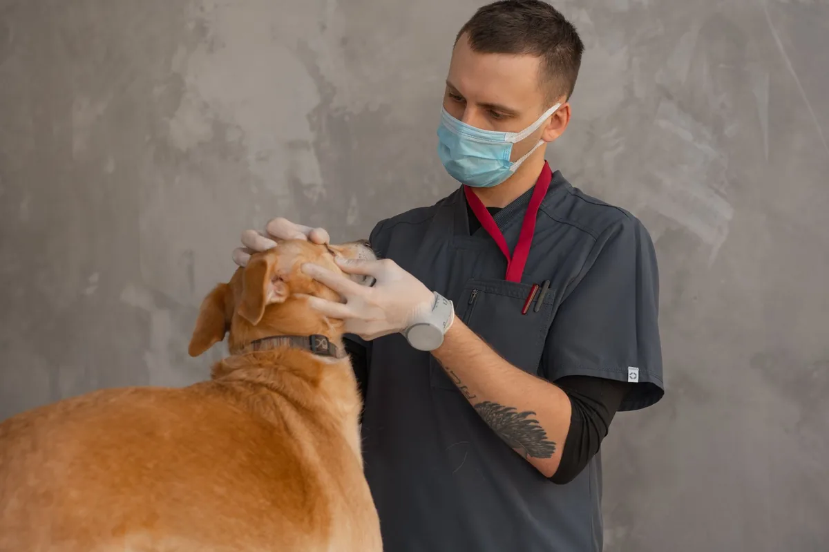
- History and physical exam: Your vet will ask about recent injuries, dental problems, or foreign‑body exposures and will palpate the lesion for firmness, pain, and discharge.
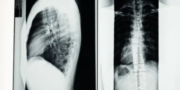
- Imaging: Radiographs (X‑rays) reveal bone involvement or deep tissue masses. Ultrasound can help assess fluid collections, while CT or MRI may be used for complex cranial or thoracic lesions (AAHA imaging guidelines, 2023).
- Fine‑needle aspirate (FNA) or biopsy: A thin needle draws cells and fluid from the mass. The sample is examined under a microscope for characteristic sulfur granules and gram‑positive filamentous bacteria.
- Culture and sensitivity: The aspirated material is cultured in an anaerobic environment to grow Actinomyces. Sensitivity testing tells the vet which antibiotics will be most effective.
- Blood work: CBC and chemistry panels check for systemic inflammation, anemia, or organ involvement that may influence treatment choices.
Because actinomycosis can mimic neoplasia (cancer) or other bacterial infections, a definitive diagnosis often hinges on the presence of filamentous organisms in the tissue sample and a positive anaerobic culture.
Treatment options
Medical treatment
Antibiotics are the cornerstone of therapy. Actinomyces species are generally susceptible to high‑dose penicillins (e.g., ampicillin, amoxicillin‑clavulanate) and tetracyclines. The typical regimen lasts 6–12 weeks, sometimes followed by a lower‑dose maintenance phase to prevent recurrence. Your vet may also prescribe:
- Clindamycin – an alternative for dogs with penicillin allergy.
- Metronidazole – useful when mixed anaerobic flora are present.
- Broad‑spectrum NSAIDs – to control pain and inflammation, but only under veterinary supervision.
Side effects can include gastrointestinal upset, loss of appetite, or, rarely, allergic reactions. Your vet will monitor blood work periodically to catch any liver or kidney changes early (Merck Veterinary Manual, 2022).
Supplements and supportive care
While antibiotics do the heavy lifting, certain supplements can aid recovery:
- Omega‑3 fatty acids (EPA/DHA): Anti‑inflammatory properties that may reduce tissue swelling; found in fish‑oil capsules or high‑quality kibble.
- Probiotics: Help maintain gut flora during prolonged antibiotic courses, reducing diarrhea risk.
- Vitamin C and zinc: Support collagen synthesis and wound healing, but only in recommended amounts.
Always discuss supplement choices with your vet; excessive fat or certain minerals can interfere with medication absorption.
Procedures or surgery
Surgery is considered when:
- Lesions are large, necrotic, or cause functional impairment (e.g., obstructing the airway).
- Antibiotics alone fail to shrink the mass after 4–6 weeks.
- There is extensive bone involvement that needs debridement.
Typical procedures involve excising the infected tissue, draining abscesses, and sometimes reconstructing bone with grafts. Post‑operative care includes a course of IV antibiotics (often 7–10 days) followed by oral therapy. Recovery from surgery can range from 2 weeks for simple drainage to 6–8 weeks for complex jaw reconstruction (cost calculators at PuppaDogs calculators).
Diet and nutrition
Nutrition plays a pivotal role in healing from any infection, especially one that may involve bone or soft‑tissue reconstruction. A diet that is highly digestible, moderate in protein, and rich in essential nutrients helps the immune system work efficiently while minimizing stress on the gastrointestinal tract.
Foods to favor
- High‑quality, highly digestible protein: Chicken, turkey, or white fish sources provide the amino acids needed for tissue repair without overloading the liver.
- Complex carbohydrates: Cooked sweet potatoes or pumpkin supply energy and gentle fiber.
- Omega‑3 enriched kibble or fish‑oil supplements: Helps control inflammation around the healing wound.
- Prescription renal or recovery diets: Formulated with controlled phosphorus and added antioxidants; useful if the infection affected the kidneys or if the dog needs a low‑fat profile.
Foods to limit or avoid
- High‑fat treats: Can exacerbate gastrointestinal upset during antibiotic therapy.
- Raw bones: While some owners feed raw, sharp bone fragments may cause additional trauma to an already compromised area.
- Excessive calcium supplements: May interfere with antibiotic absorption.
| Do feed | Limit | Avoid |
|---|---|---|
| Lean cooked meats, boiled rice, pumpkin, fish oil | Commercial treats, high‑fat foods | Raw bones, large raw meat chunks, excessive calcium |
When switching foods, do it gradually over 5–7 days to prevent digestive upset. Offer small, frequent meals (3–4 times daily) rather than one large meal, especially if the dog has a sore mouth or jaw.
Hydration is equally important. Fresh water should be available at all times, and you may add low‑salt broth to encourage drinking if your dog is reluctant.
For dogs with severe oral lesions, a soft or moistened diet (e.g., canned food mixed with water or a homemade chicken‑and‑rice mash) makes eating painless. Your vet may also recommend a prescription therapeutic diet like the “Recovery Formula” from a reputable brand, which balances protein, fat, and fiber for optimal healing.
Cost and prognosis
Because actinomycosis is rare, exact cost figures vary by region and clinic. Below is a typical breakdown for the United States and United Kingdom (costs are estimates, based on 2023 data from veterinary practice surveys):
| Item | US (USD) | UK (GBP) |
|---|---|---|
| Initial exam & blood work | $120–$200 | £80–£130 |
| Radiographs (2‑view) | $80–$150 | £60–£110 |
| FNA/biopsy & culture | $150–$300 | £100–£200 |
| Antibiotic course (12 weeks) | $250–$500 | £150–£300 |
| Surgical excision (if needed) | $2,500–$4,500 | £1,800–£3,200 |
| Post‑op hospitalization (IV antibiotics) | $1,000–$1,800 | £700–£1,200 |
Overall, owners can expect to spend anywhere from $800 to $5,500 in the US, depending on whether surgery is required. The prognosis is generally good when the infection is caught early and treated aggressively: most dogs regain normal function within 2–3 months after completing antibiotics. Chronic or extensive bone involvement can prolong recovery and may lead to residual functional deficits.
Prevention and home care
Because the bacteria are part of the normal oral flora, you can’t eliminate them entirely, but you can reduce the chance that they cause disease:
- Maintain dental health: Brush your dog’s teeth at least weekly with a dog‑safe toothpaste, and schedule professional cleanings as recommended by your vet (AAHA dental health guidelines, 2022).
- Prompt wound care: Clean any cuts, punctures, or abrasions with mild antiseptic solution and keep them covered until they heal.
- Monitor for foreign bodies: After walks in tall grass or brush, check paws and muzzle for splinters or grass awns.
- Regular vet check‑ups: Annual exams help catch early dental disease or skin infections before they become chronic.
- Balanced nutrition: A diet that supports immune function (adequate vitamins, minerals, omega‑3s) reduces susceptibility to opportunistic infections.
If your dog has a known risk factor—such as a recent dental extraction or a deep wound—ask your vet about a short prophylactic course of antibiotics. Early intervention can prevent the bacteria from establishing the thick, fibrous lesions that characterize actinomycosis.
From our vet team: “Actinomycosis can look frightening on the outside, but it’s often a manageable infection with patience, good hygiene, and a full course of antibiotics. The biggest mistake owners make is stopping treatment early because the lump seems to shrink—that’s when the bacteria can rebound. Stick with the plan, keep your dog’s mouth clean, and you’ll likely see a full recovery.”
Key takeaways
- Actinomycosis is a slow‑growing bacterial infection that creates firm, sometimes pus‑filled lesions, most often around the mouth or skin.
- Early signs include a painless swelling, reduced appetite, and foul‑smelling discharge; seek veterinary care as soon as you notice a new lump.
- Diagnosis relies on imaging, fine‑needle aspiration, and anaerobic culture; treatment is long‑term antibiotics, with surgery reserved for large or unresponsive masses.
- Recovery can take 6–12 weeks of antibiotics plus any additional time needed after surgery; costs range from a few hundred to several thousand dollars depending on complexity.
- Prevent infection by maintaining dental hygiene, promptly cleaning wounds, and avoiding prolonged exposure to foreign bodies.
Myth vs. fact
Myth: Actinomycosis always requires surgery.
Fact: Most cases respond to antibiotics alone; surgery is only needed for large, necrotic, or unresponsive lesions.
Myth: The infection is highly contagious to other pets.
Fact: Actinomycosis is not considered contagious; it arises from the dog’s own bacteria entering a wound.
Myth: A short course of antibiotics will cure the infection.
Fact: Treatment usually lasts 6–12 weeks; stopping early can lead to recurrence.
Frequently asked questions
What does actinomycosis look like in a dog?
It typically appears as a firm, sometimes ulcerated lump near the mouth, jaw, or limbs, often with a yellowish “sulfur granule” discharge.
How can I tell if my dog has actinomycosis?
If your dog has a slowly enlarging swelling, foul‑smelling discharge, and reduced appetite, a vet will perform imaging and a fine‑needle aspirate to confirm the bacterial infection.
What tests are used to confirm actinomycosis in dogs?
Diagnosis combines radiographs or CT scans, blood work, and a tissue sample sent for anaerobic culture and microscopic identification of filamentous bacteria.
Is surgery always required for canine actinomycosis?
No. Surgery is only recommended when the lesion is large, necrotic, or does not shrink with antibiotics. Many dogs heal completely with medical therapy alone.
Can antibiotics cure actinomycosis in dogs?
Yes, most dogs respond to a prolonged course of high‑dose penicillin‑type antibiotics; the treatment typically lasts 6–12 weeks and may be followed by a maintenance dose.
How can I prevent actinomycosis in my dog?
Good dental care, prompt wound cleaning, regular vet check‑ups, and monitoring for foreign bodies are the best ways to keep the bacteria from entering deeper tissues.
Ask the PuppaDogs community
Have a question this article didn’t fully answer? Want to compare notes with other dog owners who’ve been through this? Our community forum is moderated by experienced owners and vets — and answers tend to come fast. Ask in the PuppaDogs community →
References
- American College of Veterinary Internal Medicine (ACVIM). “Infectious Diseases in Dogs: Actinomycosis Overview.” 2023.
- American Animal Hospital Association (AAHA). “Dental Health Guidelines for Dogs.” 2022.
- Merck Veterinary Manual. “Actinomycosis – Bacterial Infections.” Updated 2022.
- Cornell University College of Veterinary Medicine. “Case Reports of Canine Actinomycosis.” 2022.
- World Small Animal Veterinary Association (WSAVA). “Guidelines for Antimicrobial Use in Companion Animals.” 2023.
- American Veterinary Medical Association (AVMA). “Pain Management and NSAID Use in Dogs.” 2021.
- AAHA. “Imaging Recommendations for Soft‑Tissue Infections.” 2023.
- Veterinary Oncology Group. “Differential Diagnosis of Canine Jaw Masses.” 2021.
