


Quick take: Bile duct obstruction in dogs is a blockage of the tiny tubes that carry bile from the liver to the intestine. It can cause jaundice, pain, and serious illness, but early detection and appropriate treatment—medical, surgical, or both—often lead to a good recovery. If your dog shows pale gums, a yellow tinge to the skin, or vomiting, contact your veterinarian right away.
It’s 9 p.m., and you’re sitting on the couch while your Golden Retriever, Max, usually greets you at the door, is curled up and barely moving. You notice his gums look pink‑white instead of the usual bright red, and a faint yellow hue appears on his nose and the whites of his eyes. Your heart races, you Google “yellow dog gums,” and the term “bile duct obstruction” pops up. You wonder: is this a life‑threatening emergency, and what can you do tonight?
First, take a breath. You’re not alone—many owners notice these signs just as you have. Bile duct obstruction, while not common, is a treatable condition when caught early. In the next few minutes we’ll explain what it is, why it happens, what to watch for, how vets diagnose it, and the options you have for treatment, cost, and long‑term care. We’ll also share diet tips to help Max recover comfortably.
What is bile duct obstruction in dogs?
The bile duct system is a network of tiny tubes that transports bile—a greenish fluid made by the liver—to the small intestine. Bile helps digest fats and carries waste products, including bilirubin, out of the body. When a blockage forms anywhere along this pathway, bile backs up into the liver, leading to inflammation, jaundice (yellowing of the skin and eyes), and sometimes severe infection.
Obstruction is relatively uncommon, affecting roughly 0.1–0.3 % of dogs seen in general practice, according to the American College of Veterinary Internal Medicine (ACVIM). However, certain breeds and underlying health problems make it more likely. The condition can develop suddenly (acute) or over weeks (chronic), and the clinical picture varies widely.
What causes it?
- Gallstones (cholelithiasis): Hardened bile salts can form stones that lodge in the cystic or common bile duct.
- Neoplasia: Tumors of the liver, gallbladder, or pancreas can compress or invade the ducts.
- Inflammation: Chronic pancreatitis or severe hepatitis can cause swelling that narrows the duct lumen.
- Parasites: Liver fluke (Fasciola hepatica) infections are a known cause in some regions.
- Congenital anomalies: Some puppies are born with an abnormally narrow duct or duplicate ducts that predispose to blockage.
- Breed predisposition: Shetland Sheepdogs, Miniature Schnauzers, and Cocker Spaniels have been reported more often in case series (AAHA 2022). Mixed‑breed dogs can be affected too.
Signs and symptoms
| Severity | Typical signs |
|---|---|
| Mild | Loss of appetite, occasional vomiting, slight fatigue, pale gums. |
| Moderate | Persistent vomiting (often bile‑colored), noticeable jaundice of gums, eyes, or skin, abdominal pain, fever. |
| Severe | Marked lethargy, inability to stand, severe abdominal distension, dark urine, rapid weight loss, shock. |
Early signs can be subtle—your dog might just be less enthusiastic about walks. As the blockage worsens, the classic yellowing (jaundice) becomes apparent, and vomiting may turn greenish‑brown because of bile. Pain is often felt in the right upper abdomen, and some owners notice their pet “hunches” after meals.
When to call your vet
- Call today: Pale or yellow gums, reduced appetite, occasional vomiting, mild abdominal discomfort, or any change in stool color.
- Go to an emergency clinic now: Persistent vomiting of bile, severe abdominal pain, inability to stand, rapid breathing, or signs of shock (cold paws, weak pulse).
This article is for informational purposes only and does not replace a hands‑on examination by a qualified veterinarian.
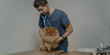
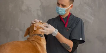
How vets diagnose it
History and physical exam. Your vet will ask about the onset of symptoms, diet, medications, and any recent illnesses. Palpation of the abdomen may reveal tenderness or a distended gallbladder.
Blood work. A complete blood count (CBC) and chemistry panel check liver enzymes (ALT, ALP, GGT), bilirubin levels, and signs of infection. Elevated bilirubin with a normal red blood cell count points to a biliary issue rather than hemolytic anemia.
Imaging. Abdominal ultrasound is the gold standard; it can visualize stones, gallbladder wall thickening, and duct dilation. Radiographs (X‑rays) may show an enlarged liver or gallbladder. In complex cases, a CT scan or magnetic resonance cholangiography provides detailed views.
Specialized tests. If infection is suspected, a bile culture can identify bacteria. In some cases, a fine‑needle aspirate of the liver or gallbladder is performed to rule out cancer. Endoscopic cholangiography—injecting contrast into the duct via a tiny scope—helps map the exact blockage location.
Treatment options
Medical treatment
When the blockage is partial or caused by inflammation, many dogs improve with supportive medicines. Commonly used drugs include:
- Ursodeoxycholic acid (a bile acid that can improve flow and protect liver cells).
- Antibiotics such as amoxicillin‑clavulanate to treat secondary bacterial infections.
- Anti‑emetics like maropitant or ondansetron to control vomiting.
- Non‑steroidal anti‑inflammatory drugs (NSAIDs) for pain, provided liver function is stable.
These medications are tailored to your dog’s weight and severity, so always ask your vet about the best option for Max.
Supplements and supportive care
Adjunctive supplements can aid liver recovery, though they are not a cure:
- Omega‑3 fatty acids (EPA/DHA): Reduce inflammation and support cell membranes.
- S‑adenosyl‑methionine (SAMe) and milk thistle (silymarin): Provide antioxidant protection for liver cells.
- Probiotics: Help maintain gut health, especially after antibiotics.
Introduce any supplement only after discussing it with your vet, as some can interact with prescribed drugs.
Procedures or surgery
If the duct is completely blocked by a stone or tumor, surgery is often required. Options include:
- Cholecystectomy: Removal of the gallbladder, which relieves pressure and prevents recurrent blockage.
- Biliary stent placement: A tiny tube inserted endoscopically to keep the duct open.
- Percutaneous drainage: Needle‑guided removal of bile to reduce inflammation before definitive surgery.
Recovery from surgery typically involves a 2–3 day hospital stay, followed by a few weeks of restricted activity. Most dogs tolerate the procedures well, but anesthesia risk is higher in dogs with severe liver disease.

Diet and nutrition
Nutrition plays a pivotal role in both recovery and prevention of bile duct problems. The goal is to give the liver a break while still providing enough calories to heal.
Key principles:
- Low‑fat, highly digestible protein: Fat stimulates bile production, so a diet with 10–12 % fat and 20–25 % high‑quality protein (e.g., chicken, turkey, or white fish) reduces workload on the biliary system.
- Moderate‑calorie density: Over‑feeding can lead to fatty liver disease, which worsens bile flow. Aim for 90–110 % of the maintenance energy requirement (MER) until the liver normalizes, then adjust based on weight.
- Added antioxidants: Vitamin E, selenium, and omega‑3 fatty acids help protect liver cells from oxidative stress.
- Prescription liver diets: Commercial therapeutic foods (e.g., “renal‑support” style formulas) are formulated with reduced copper, balanced B‑vitamins, and pre‑biotic fibers. Your vet can recommend a specific brand without bias.
When transitioning to a new diet, mix 25 % fresh food with 75 % therapeutic kibble for the first 2–3 days, then gradually increase the therapeutic portion. This helps avoid gastrointestinal upset.
| Do feed | Limit | Avoid |
|---|---|---|
| Lean boiled chicken, white fish, low‑fat cottage cheese | Cooked eggs, small amounts of healthy oil | Fatty meats, fried foods, dairy with high fat |
| Prescription liver diet (any reputable brand) | High‑protein treats (e.g., jerky) | Table scraps, pork, lamb, organ meats |
| Omega‑3 supplement (fish oil) as directed | Raw bone meals | Grains high in phosphorus (e.g., wheat, corn) |
Hydration is also essential. Offer fresh water at all times and consider adding a splash of low‑sodium broth to encourage intake. For dogs who tolerate it, a small amount of canned pumpkin (unsweetened) can aid digestion.
In the weeks following surgery, continue the low‑fat diet for at least 6–8 weeks. Your vet may gradually re‑introduce moderate‑fat foods once liver enzymes return to normal, typically confirmed by repeat blood work.
Cost and prognosis
| Item | US estimate | UK estimate |
|---|---|---|
| Initial blood work & ultrasound | $250–$600 | £150–£350 |
| Medical management (drugs 7‑10 days) | $150–$350 | £100–£250 |
| Cholecystectomy (surgery + hospital stay) | $3,000–$6,500 | £2,200–£4,500 |
| Post‑operative follow‑up (rechecks, labs) | $200–$400 | £120–£250 |
Prognosis depends on the underlying cause, how quickly treatment begins, and the dog’s overall health. Dogs with a simple gallstone blockage that are treated within a few days have a survival rate of 80–90 % and often return to normal activity within 2–3 months. Those with malignant tumors or severe liver damage have a more guarded outlook, with median survival ranging from weeks to several months.
Factors that improve outcome include early detection, stable liver enzyme values at the start of therapy, and diligent post‑operative care.
Prevention and home care
- Maintain a healthy weight: Obesity increases the risk of gallstone formation. Use the PuppaDogs calorie calculator to keep your dog at an ideal body condition.
- Regular vet check‑ups: Annual blood panels can catch rising liver enzymes before a blockage forms.
- Parasite control: Year‑round deworming for liver fluke (especially in grazing dogs) reduces one cause of blockage. Talk to your vet about appropriate products for your region.
- Balanced diet: Feed a low‑fat, high‑quality diet and avoid sudden changes that can upset bile flow.
- Monitor for early signs: Keep an eye on gum color, stool consistency, and appetite. A quick visual check of your dog’s gums each morning can catch subtle pallor early.
If your dog has had a previous obstruction, your vet may recommend periodic ultrasounds and liver enzyme monitoring to catch recurrence as early as possible.
From our vet team: “Owners often feel helpless when they see jaundice, but most cases of bile duct obstruction are manageable with prompt care. The biggest difference you can make is catching the signs early and staying in close contact with your veterinarian throughout treatment and recovery.”
Key takeaways
- Bile duct obstruction blocks bile flow, leading to jaundice, vomiting, and abdominal pain.
- Common causes include gallstones, tumors, pancreatitis, parasites, and certain breed predispositions.
- Call your vet today for pale gums or vomiting; seek emergency care if your dog can’t stand or shows signs of shock.
- Diagnosis relies on blood work, ultrasound, and sometimes advanced imaging or bile cultures.
- Treatment may be medical (meds and supplements) or surgical (gallbladder removal or stenting), with costs ranging from a few hundred to several thousand dollars.
- Low‑fat, highly digestible diets, regular vet visits, and weight control are key to preventing recurrence.
Myth vs. fact
Myth: “If my dog is jaundiced, it’s always a liver tumor.”
Fact: Jaundice can result from many reversible causes, including gallstones and inflammation. Only a thorough work‑up can determine the exact cause.
Myth: “Surgery is the only cure for a blocked bile duct.”
Fact: Partial obstructions or inflammation can often be managed medically with drugs, diet, and supportive care, especially when caught early.
Myth: “Dogs recover without any special diet.”
Fact: A low‑fat, liver‑supportive diet speeds healing and reduces the risk of a second blockage.
Frequently asked questions
What does bile duct obstruction look like in a dog?
Typical visual clues are pale or yellow‑tinged gums, yellowing of the whites of the eyes (sclera), and a greenish‑brown vomit. Your dog may also be lethargic and reluctant to eat.
How can I tell if my dog has a blocked bile duct?
The quickest home check is to look at your dog’s gum color—normal gums are bright pink. If they appear white or yellow, contact your vet immediately for blood work and an ultrasound.
What diagnostic tests are needed for bile duct obstruction?
Vets start with a CBC and chemistry panel, then use abdominal ultrasound as the primary imaging tool. In complex cases, CT scans, bile cultures, or endoscopic cholangiography may be recommended.
What are the treatment options for a dog with a blocked bile duct?
Options range from medical management with ursodeoxycholic acid, antibiotics, and anti‑emetics, to surgical removal of the gallbladder or placement of a biliary stent. The choice depends on the blockage’s cause and severity.
Is surgery always required for bile duct obstruction in dogs?
No. If the obstruction is partial or caused by inflammation, many dogs improve with medication and dietary changes. Surgery is reserved for complete blockages, gallstones, or tumors.
How much does treatment for bile duct obstruction typically cost?
Initial diagnostics (blood work and ultrasound) run $250–$600 in the U.S. Medical management adds $150–$350, while surgery can cost $3,000–$6,500. Follow‑up visits and labs are an additional $200–$400.
Ask the PuppaDogs community
Have a question this article didn’t fully answer? Want to compare notes with other dog owners who’ve been through this? Our community forum is moderated by experienced owners and vets — and answers tend to come fast. Ask in the PuppaDogs community →
References
- American College of Veterinary Internal Medicine (ACVIM). “Biliary Disease in Dogs.” 2022.
- American Animal Hospital Association (AAHA). “Canine Hepatobiliary Guidelines.” 2023.
- Merck Veterinary Manual. “Bile Duct Obstruction (Biliary Obstruction) in Dogs.” Updated 2022.
- Veterinary Clinics of North America: Small Animal Practice. “Gallstone disease in dogs.” 2021.
- World Small Animal Veterinary Association (WSAVA). “Liver Disease Consensus Statement.” 2020.
- Cornell University College of Veterinary Medicine. “Canine Liver Disease Overview.” 2023.
- American Veterinary Medical Association (AVMA). “Pet Owner’s Guide to Liver Health.” 2022.
- UK Veterinary Association. “Biliary Obstruction in Dogs – Clinical Management.” 2021.
