












Quick take: Bile leakage into the abdomen (bile peritonitis) is a medical emergency that causes severe abdominal inflammation. Dogs usually need urgent surgery or intensive medical care, and survival depends on how quickly treatment starts. Expect a hospital stay, post‑op monitoring, and a special diet during recovery.
It’s 11 p.m., and you’ve just noticed your 7‑year‑old mixed‑breed Labrador isn’t greeting you at the door. Instead, he lies on the floor, his belly looks slightly swollen, and his gums are a pale yellow‑green. Your heart races as you grab your phone and type “bile in abdomen dog” into the search bar. The answer you find—bile peritonitis—sounds terrifying, but you need to know what’s really happening, how urgent it is, and what you can do right now.
We’ll walk you through everything you need to understand: what bile peritonitis is, why it occurs, the warning signs, how vets confirm the diagnosis, treatment pathways, expected costs, diet tips for recovery, and how to help prevent a repeat episode. By the end, you’ll feel equipped to talk confidently with your veterinarian and make informed decisions for your dog’s health.
What is inflammation of the abdomen due to bile leakage in dogs?
Bile peritonitis, medically termed chemical peritonitis from bile, occurs when bile—normally stored in the gallbladder and released into the small intestine—escapes into the abdominal cavity. Bile is a digestive fluid that contains bile acids, cholesterol, and bilirubin; when it spills into the sterile peritoneal space, it triggers a severe inflammatory reaction. The abdomen becomes painful, fluid‑filled, and can quickly become infected.
Although any dog can develop this condition, it is relatively uncommon. Veterinary surveys (AAHA 2022 large‑animal practice data) estimate an incidence of less than 0.5 % of all emergency presentations, but the condition carries a high mortality risk if not treated promptly.
What causes bile leakage in dogs?
Bile can leak into the abdomen for several reasons. The most common mechanisms include:
- Gallbladder rupture – often due to a gallstone (cholelithiasis), a tumor, or severe inflammation (cholecystitis).
- Trauma – blunt force injuries from car accidents, falls, or being struck by a heavy object can tear the gallbladder or bile ducts.
- Surgical complications – accidental incision of the bile duct during spay/neuter, liver biopsy, or other abdominal procedures.
- Congenital anomalies – rare developmental defects such as biliary atresia that weaken the duct walls.
- Severe pancreatitis – inflammation of the pancreas can extend to the bile duct, causing leakage.
Breed predispositions are modest but worth noting: large, deep‑chested breeds like German Shepherds, Labrador Retrievers, and Boxers have been reported more often with gallbladder disease, while toy breeds may be more prone to traumatic injury due to their small size.
Signs and symptoms
Early detection hinges on recognizing subtle changes before the inflammation spirals. Below is a progression guide:
| Stage | Typical signs |
|---|---|
| Mild | Decreased appetite, slight abdominal bloating, low‑grade fever, occasional vomiting. |
| Moderate | Noticeable abdominal pain (dog cries when touched), lethargy, yellow‑tinged gums, frequent vomiting or diarrhea. |
| Severe | Severe abdominal distention, shock signs (rapid heartbeat, pale gums, cold limbs), inability to stand, collapse. |
Red‑flag symptoms that demand immediate veterinary attention include:
- Sudden, intense abdominal pain or “guarding” behavior.
- Pale, yellow, or grayish gums.
- Rapid breathing or heart rate.
- Vomiting bile‑colored fluid (greenish‑yellow).
- Collapse or inability to rise.
When to call your vet
Call your regular veterinarian today if you notice any of the mild‑to‑moderate signs listed above—especially loss of appetite, vomiting, or a swollen abdomen. Early evaluation can lead to diagnostic imaging before the condition worsens.
Go to an emergency veterinary hospital right now if your dog shows severe signs: severe pain, pale or yellow gums, rapid breathing, or collapse. These are signs of septic shock and require immediate fluid therapy, pain control, and possibly surgery.
Disclaimer: This article provides general information and is not a substitute for professional veterinary care. If you suspect bile peritonitis, seek veterinary help immediately.
How vets diagnose bile peritonitis
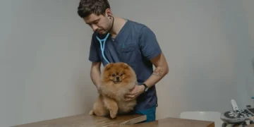
Diagnosing bile peritonitis involves a combination of history, physical exam, and targeted tests:
- Physical exam – The vet palpates the abdomen for pain, swelling, and fluid wave.
- Bloodwork – A complete blood count (CBC) often shows neutrophilic leukocytosis (high white blood cells) and elevated liver enzymes (ALT, ALP). Bile acids may be markedly increased.
- Abdominal ultrasound – The most sensitive imaging tool; it can reveal free fluid, gallbladder wall rupture, or gallstones. According to the ACVIM imaging guidelines (2023), ultrasound detects bile leakage in >90 % of cases.
- Diagnostic peritoneal lavage (DPL) or abdominocentesis – A needle draws abdominal fluid. Fluid analysis shows a high bilirubin concentration (often >5 mg/dL) compared to serum, confirming bile presence.
- CT scan (optional) – Provides detailed anatomy and can help plan surgery if ultrasound is inconclusive.
Treatment options
Medical treatment
When surgery isn’t immediately possible, supportive medical care aims to control inflammation and prevent infection:
- Broad‑spectrum antibiotics – Drugs such as amoxicillin‑clavulanic acid or enrofloxacin are commonly used to cover Gram‑negative and anaerobic bacteria.
- Anti‑inflammatory drugs – Steroids (e.g., prednisolone) or non‑steroidal anti‑inflammatory drugs (NSAIDs) may be prescribed to reduce the inflammatory cascade.
- Fluid therapy – Intravenous crystalloids restore blood pressure and correct dehydration.
- Pain management – Opioids like buprenorphine provide rapid analgesia.
- Ask your vet about these options; dosages are weight‑based and must be tailored to your dog’s condition.
Supplements and supportive care
While supplements can’t replace medical treatment, they may help the liver recover and support overall healing:
- S‑adenosyl‑methionine (SAMe) – An antioxidant that supports liver cell regeneration; often used after hepatic injury.
- Milk thistle (silymarin) – Another hepatoprotective herb; evidence from the Merck Veterinary Manual suggests modest benefits.
- Omega‑3 fatty acids (EPA/DHA) – Reduce systemic inflammation and can be added as a fish‑oil supplement once the acute phase subsides.
- These supplements should be introduced only after your vet confirms the dog is stable.
Procedures or surgery
Emergency surgery is the definitive treatment for most cases of bile leakage:
- Exploratory laparotomy – The surgeon opens the abdomen, locates the source of leakage, and either repairs the gallbladder wall, removes a ruptured gallbladder (cholecystectomy), or sutures a damaged bile duct.
- Peritoneal lavage – The abdominal cavity is flushed with sterile saline to remove bile and reduce inflammation.
- Drain placement – Temporary drains may be left in place to allow ongoing fluid removal.
- Post‑operative care includes intensive monitoring, IV fluids, pain control, and antibiotics for 5–7 days.
Diet and nutrition
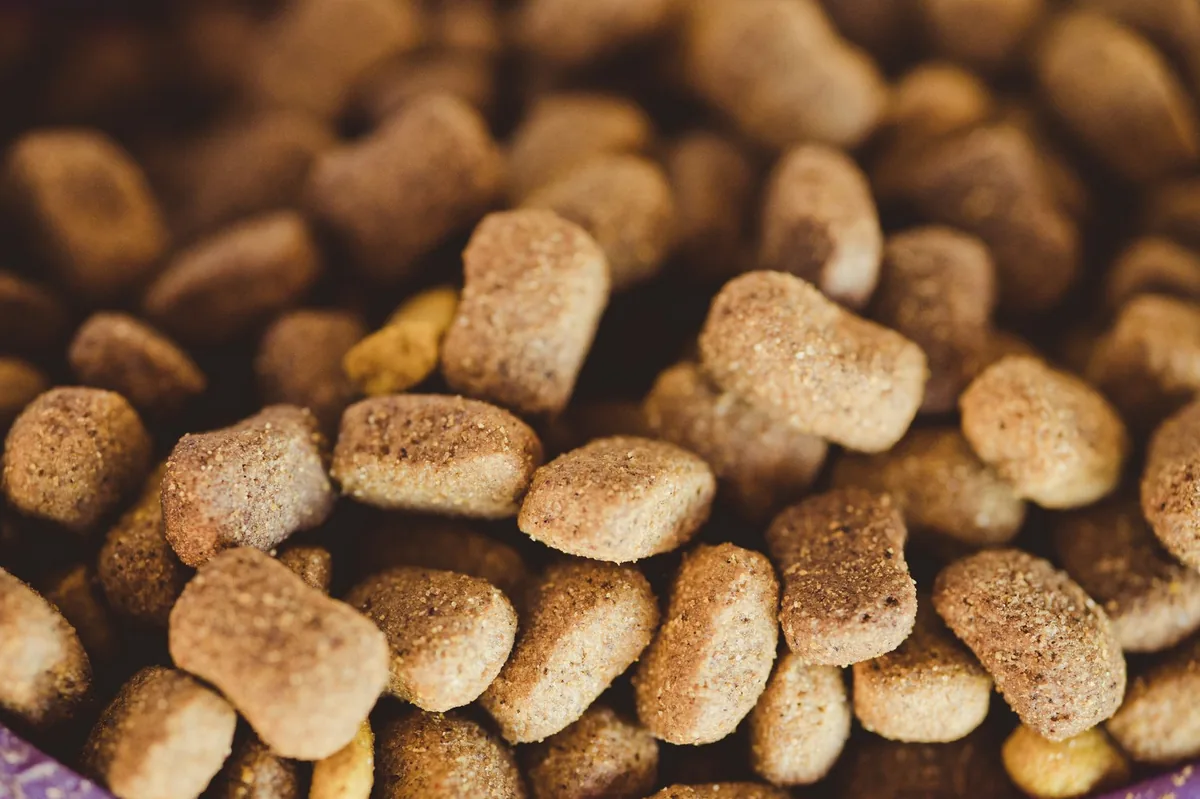
Nutrition plays a central role in recovery because the liver and gallbladder are directly involved in processing fats. The goal is to provide highly digestible, low‑fat meals that lessen bile demand while still delivering essential nutrients.
| Category | Recommendations |
|---|---|
| Do feed | Cooked lean chicken or turkey (no skin), white rice or sweet potato, low‑fat cottage cheese, boiled carrots or green beans. |
| Limit | Eggs, low‑fat fish, low‑fat commercial “sensitive stomach” kibble (if protein is high). |
| Avoid | High‑fat foods (bacon, cheese, fatty cuts), raw organ meats, dairy with high fat, treats with added oils. |
For dogs that tolerate it, a therapeutic “renal‑style” diet (moderate protein, low phosphorus, and reduced fat) can be beneficial because it eases the liver’s workload. Prescription diets such as Hill’s Prescription Diet b/d or Royal Canin Gastrointestinal Low Fat are formulated for dogs recovering from biliary or hepatic disease. Discuss with your vet which brand best matches your dog’s needs; we avoid naming a specific brand to keep the advice neutral.
Feeding frequency should be adjusted to smaller, more frequent meals (3–4 times daily) to avoid overloading the digestive tract. Start with a bland diet for 48‑72 hours, then gradually transition back to the regular diet over a week. The transition can be done by mixing 25 % new food with 75 % old food on day 1, increasing the new food by 25 % each day.
Hydration is crucial. Offer fresh water at all times, and consider adding a low‑sodium broth to encourage fluid intake if your dog is reluctant to drink.
While your dog is on medication, avoid giving any over‑the‑counter supplements without veterinary approval, as some can interfere with liver metabolism.
Cost and prognosis
Financial considerations are an important part of planning care. Below are typical cost ranges based on 2023 US and UK veterinary practice data (AAHA and VCA Hospital surveys):
| Service | US (USD) | UK (GBP) |
|---|---|---|
| Emergency ER visit (initial exam, bloodwork, imaging) | $300–$800 | £250–£600 |
| Abdominal ultrasound | $250–$500 | £200–£450 |
| Diagnostic peritoneal tap & fluid analysis | $150–$300 | £120–£280 |
| Emergency exploratory surgery (including anesthesia, meds, 24‑hr ICU) | $5,000–$9,000 | £4,000–£7,500 |
| Post‑op hospitalization (per day) | $200–$400 | £150–£350 |
| Follow‑up visits and labs (first month) | $200–$400 | £150–£300 |
Prognosis varies widely. Dogs that receive surgery within 12 hours of onset have survival rates of 60–80 % (ACVIM 2022). Those treated medically alone have lower survival (30–50 %) because bile is a potent irritant. Factors that improve outlook include younger age, absence of concurrent organ failure, and rapid stabilization of blood pressure.
Prevention and home care
Because many cases stem from gallbladder disease or trauma, the best prevention is proactive health monitoring:
- Regular wellness exams – Annual blood panels can catch early liver enzyme changes.
- Maintain a healthy weight – Obesity predisposes dogs to gallstones and biliary sludge.
- Balanced diet – Low‑fat, high‑quality protein diets reduce bile overload.
- Safe environment – Prevent falls, car‑seat injuries, and exposure to sharp objects.
- Prompt attention to GI signs – Any sudden vomiting, especially bile‑colored, warrants a vet call.
If your dog has already undergone surgery, follow these home‑care steps:
- Keep the incision clean and dry; check for swelling or discharge daily.
- Administer all prescribed medications exactly as directed.
- Offer small, frequent meals of the recommended low‑fat diet.
- Limit activity for 2–3 weeks; short leash walks are okay after the first week.
- Schedule re‑checks at the intervals your vet recommends—usually at 1 week, 2 weeks, and 1 month post‑op.
For ongoing monitoring, consider using a weight‑tracking calculator to ensure your dog stays within a healthy range.
Vet’s note
From our vet team: Bile peritonitis looks scary, but early detection and aggressive supportive care give many dogs a real chance at recovery. When you call your vet, ask about “abdominal fluid analysis” and “ultrasound for gallbladder rupture”—these tests will pinpoint the problem fast. Stay calm, bring a list of any recent injuries or surgeries, and trust that the veterinary team will tailor fluids, antibiotics, and pain meds to your dog’s size and severity.
Key takeaways
- Bile peritonitis is a medical emergency caused by bile spilling into the abdomen, often from gallbladder rupture.
- Watch for sudden abdominal pain, yellow or pale gums, and bile‑colored vomiting—these are red‑flag signs.
- Diagnosis relies on ultrasound and peritoneal fluid analysis; rapid imaging can save time.
- Emergency surgery offers the best survival odds, but intensive medical management is also possible.
- Post‑op recovery needs a low‑fat, highly digestible diet and close monitoring for infection.
- Regular wellness checks, weight control, and safe environments help prevent future bile leakage.
Myth vs. fact
Myth: Bile peritonitis can be treated at home with over‑the‑counter meds.
Fact: It requires veterinary diagnostics, IV fluids, antibiotics, and often surgery; home care is only supportive after professional treatment.
Myth: All dogs with gallbladder disease will eventually rupture.
Fact: Many dogs have gallbladder inflammation or stones without rupture; early detection and diet changes can prevent rupture.
Myth: If the dog looks “fine” after vomiting, the problem is over.
Fact: Bile is highly irritating; even mild‑looking vomiting can signal serious internal leakage that needs prompt vet evaluation.
Frequently asked questions
What causes bile to leak into a dog’s abdomen?
Bile leaks when the gallbladder wall or bile ducts are damaged, most commonly from gallbladder rupture due to gallstones, severe inflammation, trauma, or surgical mishaps.
What signs should I look for if my dog has bile peritonitis?
Key signs include sudden abdominal pain, swollen belly, pale or yellow gums, bile‑colored vomiting, rapid breathing, and weakness or collapse.
How is bile peritonitis confirmed by a veterinarian?
Veterinarians use abdominal ultrasound to locate fluid and gallbladder damage, and they often perform a peritoneal tap—analyzing the fluid’s bilirubin level, which is markedly higher than blood levels.
What are the treatment options for bile leakage in dogs?
Options range from emergency exploratory surgery to repair or remove the gallbladder, to intensive medical management with IV fluids, antibiotics, and anti‑inflammatory drugs when surgery isn’t immediately possible.
Can a dog survive bile peritonitis without surgery?
Some dogs improve with aggressive medical therapy alone, but survival rates are lower (30–50 %). Surgery within the first 12 hours offers the best chance of a full recovery.
What is the typical recovery time after bile peritonitis surgery?
Most dogs stay hospitalized for 3–5 days for monitoring, then need 2–3 weeks of restricted activity and a low‑fat diet. Full healing of the abdominal incision usually takes 4–6 weeks.
Ask the PuppaDogs community
Have a question this article didn’t fully answer? Want to compare notes with other dog owners who’ve been through this? Our community forum is moderated by experienced owners and vets — and answers tend to come fast. Ask in the PuppaDogs community →
References
- American Animal Hospital Association (AAHA) 2022 Large‑Animal Practice Guidelines.
- American College of Veterinary Internal Medicine (ACVIM) Imaging Recommendations, 2023.
- Merck Veterinary Manual, “Bile Peritonitis” entry.
- Cornell University College of Veterinary Medicine, “Gallbladder Disease in Dogs” lecture notes.
- University of California, Davis Veterinary Medicine, “Abdominal Fluid Analysis” protocol.
- World Small Animal Veterinary Association (WSAVA) Guidelines on Veterinary Emergency Care, 2022.
- Veterinary Cancer Society, “Biliary Tract Tumors” fact sheet.
- American Veterinary Medical Association (AVMA) Pet Health Insurance Cost Survey, 2023.
- Royal Veterinary College, “Dietary Management of Hepatic and Biliary Disorders” (2022).
- Pet Care Industry Association, “Cost of Emergency Surgery in the United States” report, 2023.


