












Quick take: Fluid that builds up in a dog’s lungs without a heart problem is called non‑cardiac pulmonary edema or pleural effusion. It can be caused by infection, trauma, toxins, tumors, or severe inflammation. Prompt veterinary care—often oxygen, medication, and sometimes a fluid‑draining procedure—greatly improves the odds of recovery.
It’s 11 p.m., and your 7‑year‑old mixed‑breed Labrador is lying on the couch, breathing shallowly with his mouth open. You notice his ribs are more visible than usual and his gums look a pale pink instead of the usual bright red. The coughing that started a few hours ago has turned into a wet, gurgling sound. Your heart races as you scroll through articles, wondering if this could be a heart issue or something else.
First, take a deep breath. Not all lung fluid points to a failing heart. Many dogs develop fluid in the lungs from infections, injuries, or other diseases that don’t involve the heart. Understanding what’s happening, what to look for, and how treatment works can help you act quickly and keep your companion safe.
In this guide we’ll explain what non‑cardiac fluid in the lungs is, why it happens, the signs you can spot at home, how vets find the cause, what treatments are available, typical costs, and steps you can take to prevent a recurrence. We’ll also give you a handy diet plan for the recovery period and a quick checklist for emergencies.
What is collection of fluid in the lungs (not due to heart disease) in dogs?
When fluid accumulates in a dog’s lungs without a heart‑related cause, veterinarians refer to it as non‑cardiac pulmonary edema (fluid inside the lung tissue) or pleural effusion (fluid in the space surrounding the lungs). Both conditions interfere with normal breathing, but the underlying mechanisms differ from the “congestive heart failure” scenario most owners first think of.
In pulmonary edema, the tiny air sacs (alveoli) fill with watery fluid, often because of severe inflammation, toxins, or injury. Pleural effusion, on the other hand, is fluid that gathers in the pleural cavity—the thin space between the lungs and the chest wall—usually from infection, tumors, or trauma.
These problems are relatively uncommon compared with heart‑related fluid buildup, but they are serious enough that most affected dogs need veterinary attention within hours to days. According to the American College of Veterinary Internal Medicine (ACVIM), pulmonary edema of non‑cardiac origin accounts for roughly 10‑15 % of all acute respiratory emergencies in dogs.
What causes it?
Non‑cardiac fluid can arise from several distinct disease processes. Below is a quick overview of the most frequent culprits:
| Cause | Typical mechanism | Breed or age trends |
|---|---|---|
| Pneumonia (bacterial, viral, fungal) | Inflammation and infection increase capillary permeability, leaking fluid into alveoli. | All breeds; puppies and senior dogs are most vulnerable. |
| Acute respiratory distress syndrome (ARDS) | Severe systemic inflammation (e.g., after sepsis or severe trauma) overwhelms lung vessels. | Often seen in large breeds after accidents or surgery. |
| Trauma (blunt force, penetrating wounds) | Direct damage to lung tissue or chest wall leads to leakage of blood or serous fluid. | Any dog that experiences a car accident or fall. |
| Neoplasia (lung tumors, metastatic cancer) | Tumor growth disrupts normal vasculature, causing fluid exudation. | Older dogs, especially Boxers, Golden Retrievers, and German Shepherds. |
| Toxins (inhaled smoke, certain chemicals) | Irritation of the airway lining leads to leaky capillaries. | Urban dogs or those near farms; no breed restriction. |
| Allergic or immune‑mediated reactions | Excess histamine and cytokines increase vessel permeability. | Often seen in dogs with known allergies; no specific breed pattern. |
Signs and symptoms
Because the lungs are essential for oxygen exchange, even a modest amount of fluid can cause noticeable distress. Below is a tiered chart to help you match what you see with the likely severity.
| Severity | Typical signs you may notice |
|---|---|
| Mild | Occasional coughing, slight panting after exercise, mild lethargy. |
| Moderate | Persistent cough, open‑mouth breathing, pale or bluish gums, reduced appetite, reluctance to climb stairs. |
| Severe | Rapid, labored breathing (dyspnea), cyanosis around nose or tongue, inability to lie down comfortably, collapse, frothy discharge from nose or mouth. |
Other clues include a “wet” sound when you place a hand on the chest, a “gurgling” noise during exhalation, and a noticeable increase in heart rate (often >140 bpm in a resting adult). If your dog is also vomiting, has a fever, or shows signs of pain, the underlying cause may be infection or trauma.
When to call your vet
Call your vet today (within the next 12 hours) if:
- Your dog is coughing more than a few times per hour, or the cough is wet, gagging, or producing foam.
- Breathing is noticeably faster than normal (over 30 breaths per minute at rest) or you hear labored effort.
- Gums look pale, gray, or bluish, or the tongue appears swollen.
- Your dog is lethargic, refuses food, or seems uncomfortable when lying down.
Go to an emergency veterinary hospital right now if you see any of these:
- Severe dyspnea—dog is struggling to breathe, flattens the chest, or is gasping.
- Sudden collapse, loss of consciousness, or seizures.
- Large amount of frothy discharge from nose or mouth.
- Rapid worsening within minutes (e.g., coughing turns to choking).
These signs indicate a life‑threatening emergency. Even if you’re unsure, it’s safer to err on the side of caution and seek immediate care.
This article is for informational purposes only and does not replace a hands‑on veterinary exam.
How vets diagnose it
Veterinarians follow a systematic approach to pinpoint the exact reason fluid is in the lungs.
- History and physical exam: The vet asks about recent illnesses, injuries, exposures (smoke, chemicals), vaccination status, and medications. A thorough lung and heart auscultation (listening with a stethoscope) helps differentiate fluid from heart disease.
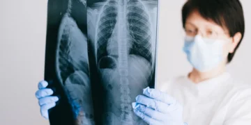
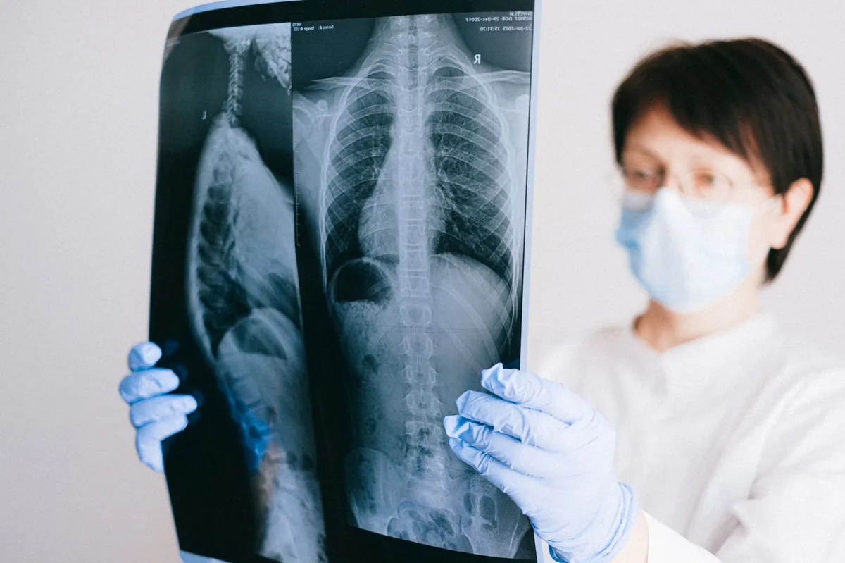
- Chest radiographs (X‑rays): The most common first‑line imaging. X‑rays reveal fluid patterns—diffuse “white‑out” for pulmonary edema or a distinct line for pleural effusion.
- Thoracic ultrasound: Provides real‑time images of fluid pockets and can guide a needle for fluid removal (thoracocentesis).
- CT scan (computed tomography): Used when the cause is unclear or a tumor is suspected; offers detailed cross‑sectional views of lung tissue.
- Fluid analysis: If fluid is tapped, it’s examined for cell type, protein content, and presence of bacteria or cancer cells. This helps distinguish infectious pus from sterile inflammation.
- Blood work: A complete blood count (CBC) checks for infection or anemia; chemistry panel evaluates organ function; arterial blood gases (ABG) assess oxygen and carbon‑dioxide levels.
- Additional tests: Depending on suspicion, the vet may run a PCR panel for respiratory pathogens, a bronchoscopy to look inside the airway, or a heart ultrasound (echocardiogram) to definitively rule out cardiac disease.
Treatment options
Medical treatment
Most dogs improve with a combination of supportive and targeted drugs. Commonly used classes include:
- Oxygen therapy: Delivered via cage‑side flow‑through mask or a nasal cannula to correct low blood oxygen.
- Diuretics: Furosemide (Lasix) helps move excess fluid out of the lungs by increasing urine production. Your vet will adjust the dose based on weight and kidney function.
- Corticosteroids: Prednisone or Dexamethasone reduce inflammation when the fluid is due to an immune reaction or severe ARDS.
- Antibiotics: If infection is suspected, drugs such as amoxicillin‑clavulanate or enrofloxacin are prescribed. The exact choice depends on culture results if fluid is sampled.
- Bronchodilators and mucolytics: Medications like theophylline or inhaled albuterol open airways and thin mucus, making breathing easier.
- Antitoxins or antivirals: For specific toxin exposures (e.g., smoke inhalation) or viral pneumonia, treatments are tailored accordingly.
All medications are administered under veterinary guidance. Ask your vet about each option, especially if your dog is already on other drugs.
Supplements and supportive care
Evidence‑based adjuncts can aid recovery, though they never replace prescription therapy.
- Omega‑3 fatty acids (EPA/DHA): Fish‑oil supplements have anti‑inflammatory properties that may lessen lung edema. Look for products formulated for dogs with a guaranteed EPA/DHA level.
- Vitamin E and selenium: Antioxidants that protect lung tissue from oxidative damage. A balanced commercial diet usually provides sufficient amounts, but a vet may suggest a supplement for severe cases.
- Probiotics: Maintaining gut health supports overall immunity, which can be valuable when fighting pneumonia.
- Hydration support: Sub‑cutaneous (SQ) fluids or IV fluids are given to keep the blood volume stable while diuretics are working.
Procedures or surgery
When fluid cannot be cleared with medication alone, or when a mass is the source, procedural intervention is required.
- Thoracocentesis: A sterile needle is inserted into the pleural space to withdraw fluid. This can provide rapid relief and also supplies the sample for laboratory analysis.
- Chest tube placement: For ongoing drainage, a small tube may be left in place for 24‑48 hours.
- Bronchoscopy: A tiny camera is passed into the airway to remove debris, collect samples, or deliver localized medication.
- Surgical removal: If a tumor or severe trauma is the culprit, surgery may be recommended. Recovery from thoracic surgery can take several weeks and often requires intensive postoperative care.
Diet and nutrition
Nutrition plays a supportive role in healing lung tissue, controlling inflammation, and preventing fluid re‑accumulation. While there is no “one‑size‑fits‑all” prescription diet specifically for pulmonary edema, the following guidelines align with recommendations from the American Veterinary Medical Association (AVMA) and the World Small Animal Veterinary Association (WSAVA).
Key principles
- Moderate sodium: Excess dietary salt can promote fluid retention. Choose commercial foods that list “low sodium” or “reduced sodium” on the label. Avoid table scraps like processed meats, cheese, and salty snacks.
- High‑quality protein: Protein supports tissue repair and immune function. Look for named protein sources (e.g., chicken, salmon) as the first ingredient.
- Easily digestible carbs: Rice, barley, or sweet potato provide energy without taxing the gastrointestinal tract.
- Omega‑3 fatty acids: As mentioned, EPA/DHA reduce inflammation. Foods formulated for “joint health” often contain added fish oil, which can be beneficial for lung recovery as well.
- Antioxidants: Vitamin E, selenium, and vitamin C help limit oxidative damage caused by inflammation. A balanced diet typically meets these needs, but a supplement can be discussed with your vet if the dog is on a limited‑ingredient or homemade diet.
Foods to favor, limit, and avoid
| Do feed | Limit | Avoid |
|---|---|---|
| Commercial dry or wet foods labeled “low sodium” or “light” | Table scraps with moderate fat (e.g., small pieces of cooked chicken without skin) | Processed meats, cheese, bacon, and salty treats |
| Cooked white rice or sweet potato mixed with lean protein | High‑fat canned foods (excess oil) | Raw bones that may splinter; high‑phosphorus diets if kidney function is compromised |
| Fish‑oil supplement (EPA/DHA 300–500 mg per day for a 20 lb dog) | Commercial treats with added salt | Any food containing onion, garlic, or chocolate |
Feeding schedule and transition tips
During the acute phase, many dogs have a reduced appetite. Offer small, frequent meals (3–4 times daily) of bland, easily digestible food such as boiled chicken and rice. Gradually re‑introduce their regular diet over 5–7 days as breathing improves.
When transitioning to a therapeutic or “light‑sodium” food, follow the “mix‑and‑match” method: start with 25 % new food + 75 % old food, then increase the new food by 25 % every two days. This helps avoid gastrointestinal upset and keeps the dog’s caloric intake stable.
Hydration is crucial. Fresh water should be available at all times. If your dog is reluctant to drink, try adding a splash of low‑sodium broth (no onion or garlic) or offering ice cubes.
Cost and prognosis
Financial considerations are an important part of any veterinary plan. Below are typical cost ranges for the United States and United Kingdom, based on data from the PuppaDogs cost estimator and AAHA guidelines. Prices vary by region, clinic, and individual case complexity.
| Service | US estimate (USD) | UK estimate (GBP) |
|---|---|---|
| Initial exam + basic blood work | $150–$300 | £120–£250 |
| Chest X‑ray (2‑view) | $100–$200 | £80–£150 |
| Thoracic ultrasound | $200–$350 | £150–£280 |
| Fluid analysis (if thoracocentesis performed) | $100–$180 | £80–£150 |
| Oxygen therapy (hospital stay 24 hrs) | $250–$500 | £200–£400 |
| Medical treatment (diuretics, steroids, antibiotics) | $100–$400 | £80–£300 |
| Thoracocentesis or chest tube placement | $300–$800 | £250–£600 |
| Surgical tumor removal (if needed) | $2,000–$5,000 | £1,500–£4,000 |
Overall prognosis depends on the underlying cause, the speed of treatment, and the dog’s age and overall health. For infections caught early, survival rates exceed 80 % with appropriate antibiotics and supportive care. Trauma‑related edema has a more variable outcome—dogs that receive rapid oxygen and fluid drainage often recover fully, while severe lung contusions may lead to chronic respiratory issues.
When the fluid is due to a malignant tumor, the outlook is guarded; however, palliative care can still provide a good quality of life for several months.
Prevention and home care
While not all cases are preventable, many risk factors can be managed.
- Vaccination: Keep core vaccines up to date (distemper, parvovirus, adenovirus, and Bordetella) to reduce the chance of bacterial or viral pneumonia.
- Safe environment: Avoid exposing your dog to heavy smoke, chemical fumes, or dusty construction sites. Use a pet‑safe air purifier if you live in a high‑pollution area.
- Post‑surgical monitoring: After anesthesia, monitor for coughing or labored breathing for at least 24 hours. Follow your vet’s instructions on pain meds, as some NSAIDs can affect kidney function and fluid balance.
- Weight management: Overweight dogs have higher risk of respiratory compromise. A balanced diet and regular exercise keep the lungs and heart healthy.
- Prompt treatment of infections: Early antibiotics for bacterial infections and timely care for upper respiratory signs can stop fluid from building up.
- Regular check‑ups: Annual exams (or semi‑annual for senior dogs) allow vets to catch early lung changes before they become critical.
At home, keep a small notebook of your dog’s breathing rate, activity tolerance, and any coughing episodes. This log helps the vet gauge progress and adjust treatment.
From our vet team: “When fluid builds up quickly, the most important thing is to get oxygen into the bloodstream as fast as possible. Even if you’re scared, staying calm and getting your dog to a clinic within a few hours dramatically improves the odds of a full recovery.”
Key takeaways
- Non‑cardiac lung fluid can arise from infection, trauma, toxins, or tumors, and is treatable when caught early.
- Watch for rapid breathing, pale gums, persistent coughing, or open‑mouth panting—these are red‑flag signs to call your vet now.
- Diagnosis typically involves chest X‑rays, ultrasound, and fluid analysis; your vet will rule out heart disease with an echocardiogram if needed.
- Treatment combines oxygen, diuretics, steroids, antibiotics, and sometimes fluid drainage or surgery.
- Use a moderate‑sodium, high‑protein diet with omega‑3 supplements during recovery; transition slowly back to regular food.
- Prevent future episodes by keeping vaccinations current, avoiding smoke and toxins, maintaining a healthy weight, and monitoring after surgery.
Myth vs. fact
Myth: All fluid in a dog’s lungs means the heart is failing.
Fact: Fluid can collect for many non‑cardiac reasons, such as pneumonia, trauma, or cancer. A proper veterinary workup distinguishes the cause.
Myth: Once the fluid is gone, the problem is solved.
Fact: Underlying disease may still be present; ongoing monitoring and treatment are needed to prevent recurrence.
Myth: Home remedies like “salt water” will dissolve lung fluid.
Fact: Only veterinary‑administered oxygen, medications, and, when needed, fluid drainage can safely resolve the condition.
Frequently asked questions
Can lung fluid in dogs be cured without surgery?
Most cases are managed medically with oxygen, diuretics, steroids, and antibiotics, and they resolve without surgery. Surgery is only needed if a tumor or persistent fluid source cannot be treated otherwise.
How quickly does non‑cardiac pulmonary edema develop in dogs?
It can develop within hours after a severe infection, trauma, or toxin exposure. Early signs may be subtle, but once labored breathing begins, the condition can become critical in less than 24 hours.
What diagnostic tests confirm fluid in the lungs of a dog?
Chest X‑rays and thoracic ultrasound are the primary imaging tools. Fluid analysis from a thoracocentesis, blood work, and sometimes a CT scan or bronchoscopy help identify the exact cause.
Are there any breeds more likely to develop lung fluid without heart problems?
Large breeds such as Boxers, Golden Retrievers, and German Shepherds have been reported more often with trauma‑related edema, while older dogs of any breed are prone to pneumonia‑induced fluid. No breed is immune.
What is the typical recovery time for a dog treated for lung fluid accumulation?
Most dogs improve within 2–5 days of starting oxygen and medication. Full recovery, including weaning off supplemental oxygen and returning to normal activity, usually takes 1–3 weeks, depending on the underlying cause.
What are the costs of treating non‑cardiac lung fluid in dogs?
Diagnostic workups range from $300 to $800, while treatment (oxygen, medications, possible thoracocentesis) adds $200–$1,000. Surgery, when required, can increase total expenses to $2,000–$5,000. Costs vary by region and clinic.
Ask the PuppaDogs community
Have a question this article didn’t fully answer? Want to compare notes with other dog owners who’ve been through this? Our community forum is moderated by experienced owners and vets — and answers tend to come fast. Ask in the PuppaDogs community →
References
- American College of Veterinary Internal Medicine (ACVIM). “Acute Respiratory Distress Syndrome in Dogs.” 2022 clinical guidelines.
- American Animal Hospital Association (AAHA). “Guidelines for Diagnosis and Treatment of Pulmonary Edema.” 2023 edition.
- Merck Veterinary Manual. “Pulmonary Edema, Non‑Cardiac.” Updated 2024.
- World Small Animal Veterinary Association (WSAVA). “Nutritional Management of Dogs with Respiratory Disease.” 2021 position statement.
- American Veterinary Medical Association (AVMA). “Vaccination Recommendations for Canine Respiratory Pathogens.” 2023.
- Veterinary Clinics of North America: Small Animal Practice. “Thoracic Ultrasound and Fluid Analysis in Dogs.” 2022.
- University of California, Davis School of Veterinary Medicine. “Diagnostic Approach to Canine Pleural Effusion.” 2023.
- British Veterinary Association (BVA). “Cost of Veterinary Care: Diagnostic and Treatment Estimates.” 2022.


