













Quick take: A pericardio‑peritoneal hernia is a rare defect where abdominal organs slip into the chest cavity through a tear between the pericardium (heart sac) and the peritoneum (abdominal lining). It can be congenital or caused by trauma, and while some dogs live with mild signs, many need prompt surgical repair. Early detection, imaging, and a skilled surgeon give the best chance for a full recovery.
It’s 11 p.m., and you’re scrolling through your phone because your 9‑year‑old mixed‑breed Labrador just stopped trotting to the kitchen and is lying low, eyes half‑closed. When you gently lift his front leg, his ribs feel oddly hollow, and his gums look a little paler than usual. Your heart races—could this be something serious?
Many owners describe that exact moment: a sudden change in energy, a subtle “thump” in the chest, or a strange “gurgle” when they listen to their dog’s breathing. The fear that something is wrong is natural, but you’re not alone. In the next few minutes we’ll explain exactly what a pericardio‑peritoneal hernia is, why it happens, what to look for, how vets diagnose and treat it, and what you can expect in terms of cost, recovery, and long‑term care.
Our goal is to give you a clear roadmap—so you know when to act, what questions to ask your vet, and how to support your dog through the healing process.
What is a hernia between the pericardium and peritoneum in dogs?
A pericardio‑peritoneal hernia (sometimes called a pericardial‑diaphragmatic hernia) occurs when a tear or defect creates a passage between the pericardium—the thin sac that surrounds the heart—and the peritoneum, the membrane lining the abdominal cavity. Through this opening, portions of the stomach, intestines, liver, or spleen can migrate upward into the chest cavity, crowding the heart and lungs.
In healthy dogs the diaphragm and pericardial membrane act as separate barriers. When that barrier is compromised, the pressure differential between the abdomen and thorax can pull soft tissue through the defect, much like a balloon slipping through a hole in a sack.
Although the condition is rare—estimated at less than 0.01 % of all canine thoraco‑abdominal presentations—it is most commonly reported in large breeds and in dogs that have experienced severe blunt trauma (e.g., car accidents) or that were born with a congenital weakness.
What causes it?
- Congenital defects: Developmental errors in the diaphragm or pericardium during fetal growth can leave a small opening that may stay silent for months or years.
- Traumatic injury: High‑impact accidents, such as being hit by a vehicle or a fall from height, can rupture the pericardial‑peritoneal interface.
- Breed predisposition: Large, deep‑chested breeds—Great Danes, German Shepherds, and Mastiffs—appear over‑represented, likely because of the greater mechanical forces across the thoraco‑abdominal diaphragm.
- Secondary factors: Chronic coughing, severe vomiting, or increased intra‑abdominal pressure (e.g., from obesity) can exacerbate a pre‑existing weak spot.
Understanding these risk factors helps you discuss preventive measures with your veterinarian, especially if your dog belongs to a high‑risk breed.
Signs and symptoms
Because the herniated organs occupy space meant for the heart and lungs, clinical signs can range from subtle to life‑threatening. Early signs often mimic other thoraco‑abdominal problems, making a thorough exam essential.
| Severity | Typical signs |
|---|---|
| Mild | Reduced stamina, occasional coughing, slight abdominal distension, mild respiratory rate increase. |
| Moderate | Persistent cough, difficulty breathing on exertion, pale gums, decreased appetite, occasional vomiting. |
| Severe | Labored breathing at rest, rapid heart rate, cyanotic gums, collapse, abdominal pain, shock. |
When to call your vet
Call your vet today if you notice any of the following:
- Persistent cough or gagging that doesn’t improve within 24 hours.
- Reduced appetite, mild vomiting, or a noticeable decrease in energy.
- Pale or bluish gums, especially after exercise.
- Abdominal swelling or a “full” feeling when you gently press the belly.
Go to an emergency veterinary hospital right now if you see any of these urgent signs:
- Severe difficulty breathing (open‑mouth breathing, rapid respiratory rate).
- Sudden collapse, unresponsiveness, or seizures.
- Bright red or dark, tarry stools indicating possible gastrointestinal bleeding.
- Shock signs: weak pulse, cold extremities, or a rapid, weak heartbeat.
These guidelines are for information only and do not replace professional care. If you’re ever unsure, err on the side of caution and contact a veterinarian.
How vets diagnose it
Diagnosis starts with a thorough history—asking about recent trauma, coughing episodes, vomiting, or changes in activity. A physical exam may reveal a muffled heart sound, abnormal lung sounds, or a palpable abdominal mass shifting with respiration.
Imaging is the cornerstone:
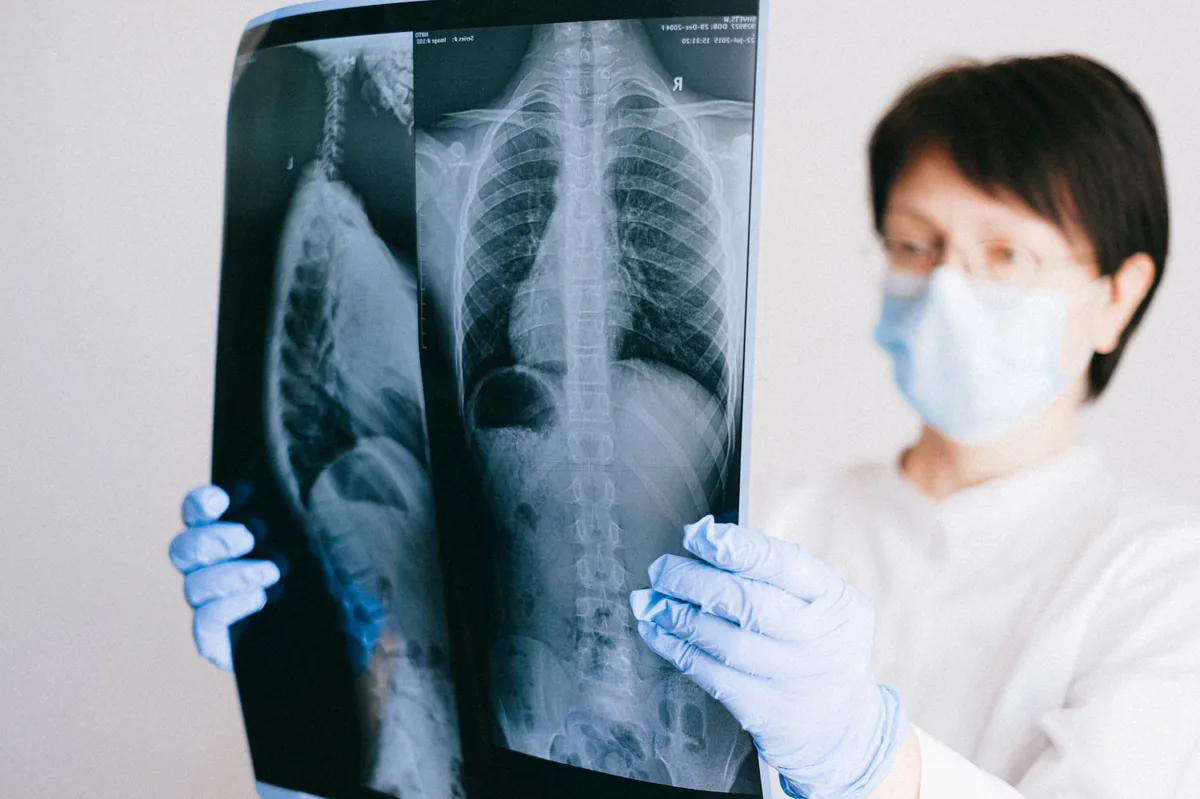
- Thoracic radiographs (X‑rays): Show abnormal silhouettes of abdominal organs within the chest cavity.
- Abdominal ultrasound: Helps identify which organ(s) have migrated and assesses blood flow.
- CT scan or MRI: Provides a 3‑dimensional view of the defect, useful for surgical planning.
- Echocardiography (heart ultrasound): Evaluates cardiac compression and ensures the heart itself is not damaged.
Blood work (CBC, chemistry panel) may reveal anemia, electrolyte imbalances, or signs of organ compromise, guiding pre‑operative stabilization.
Treatment options
Medical treatment
Medical management is usually a bridge to surgery rather than a definitive cure. It may include:
- Anti‑inflammatory drugs: NSAIDs such as carprofen or meloxicam to reduce thoracic inflammation (dose determined by your vet).
- Antibiotics: Broad‑spectrum agents like amoxicillin‑clavulanate if secondary bacterial infection is suspected.
- Fluid therapy: Intravenous crystalloids to correct dehydration or shock while preparing for surgery.
- Oxygen supplementation: To support breathing if the lungs are compromised.
All medication choices should be discussed with your veterinarian—ask your vet about these options and any potential side effects.
Supplements and supportive care
While supplements cannot close the hernia, they can aid recovery and support organ function:
- Omega‑3 fatty acids (EPA/DHA): Anti‑inflammatory benefits may help reduce post‑operative swelling.
- Vitamin E and selenium: Antioxidants that support lung tissue health.
- Probiotics: Useful if the dog experiences gastrointestinal upset after surgery.
- Glutamine: May assist intestinal healing when the stomach or intestines are involved.
Supplements should be introduced after the first post‑operative check and only under veterinary guidance.
Procedures or surgery
Definitive repair almost always requires surgery. The most common technique is a thoracotomy (an incision between the ribs) or a median sternotomy** to directly visualize the defect. The surgeon reduces the herniated organ(s) back into the abdomen, then closes the tear with non‑absorbable sutures, sometimes reinforcing it with a synthetic mesh.
Key points about surgery:
- Procedures are performed under general anesthesia with intensive monitoring (AAHA anesthesia guidelines).
- Hospital stays range from 2–5 days, depending on the dog’s age, overall health, and any complications.
- Post‑operative pain is managed with multimodal analgesia (opioids, NSAIDs, local blocks).
- Complication rates are low (< 10 %) when performed by an experienced board‑certified surgeon, but possible issues include infection, recurrence of the hernia, or pleural effusion.
Diet and nutrition
Nutrition plays a supportive role during recovery. The goal is to provide easily digestible, high‑quality protein while avoiding excessive pressure on the diaphragm and pericardium.
Key recommendations:
- Highly digestible protein: Sources like boiled chicken, turkey, or commercially available “wet” diets formulated for recovery help maintain lean body mass without over‑loading the digestive tract.
- Moderate fat: About 10‑12 % of calories from fat provides energy but reduces the risk of gastric dilation.
- Low sodium: Reduces fluid retention and eases cardiac workload; avoid table scraps, processed meats, and salty treats.
- Small, frequent meals: Feeding 4‑6 smaller meals per day limits gastric distension, which could otherwise push abdominal contents upward.
- Hydration: Fresh water should always be available; consider adding a little low‑sodium broth to encourage intake if the dog is reluctant.
- Prescription cardiac diets: If the hernia has caused cardiac compression, a diet formulated for heart health (e.g., low‑phosphorus, omega‑3 enriched) may be recommended. Discuss brand‑specific options with your vet—do not choose a brand based solely on marketing.
| Do feed | Limit | Avoid |
|---|---|---|
| Boiled lean meats, low‑fat cottage cheese, cooked sweet potatoes | Commercial treats > 5 % of daily calories | High‑salt foods, raw bones that can splinter |
| Prescription cardiac formulas (if advised) | Fatty cuts of meat | Heavy, greasy foods that cause bloating |
| Omega‑3 enriched fish oil (vet‑approved) | Large meals (> 2 cups at once) | Table scraps with onions or garlic |
Transition to any new diet gradually over 5–7 days to avoid gastrointestinal upset. If your dog is a picky eater, warm the food slightly or add a low‑sodium broth to enhance aroma.
For personalized calorie needs, you can use our dog nutrition calculator to estimate daily energy requirements based on weight, activity level, and recovery stage.
Cost and prognosis
Because the condition is uncommon, costs can vary widely. Below are typical ranges for the United States and United Kingdom, presented as estimates—not guarantees.
| Item | US (USD) | UK (GBP) |
|---|---|---|
| Initial consult & basic bloodwork | $150–$300 | £80–£150 |
| Radiographs (2‑view thoracic) | $200–$400 | £120–£250 |
| Abdominal ultrasound | $300–$600 | £180–£350 |
| CT scan (if needed) | $800–$1,200 | £600–£1,000 |
| Surgical repair (including anesthesia, surgeon fee, mesh if used) | $3,500–$6,500 | £2,500–£4,500 |
| Hospitalization (2–5 days) | $500–$1,200 | £300–£700 |
| Post‑op meds & follow‑up visits | $200–$400 | £120–£250 |
Overall, owners can expect a total expense of roughly $5,000–$9,000 in the US or £3,500–£6,000 in the UK when all diagnostics, surgery, and post‑operative care are included.
Prognosis is generally good when the hernia is repaired promptly and the dog has no underlying heart disease. Survival rates exceed 85 % for uncomplicated cases (AAHA 2022 Surgical Guidelines). Dogs that present with severe respiratory compromise or concurrent organ failure have a more guarded outlook, emphasizing the importance of early detection.
Prevention and home care
Because many cases are trauma‑related, the most effective preventive measures focus on minimizing injury and monitoring at‑risk dogs:
- Weight management: Keeping your dog at a healthy body condition reduces intra‑abdominal pressure.
- Safe environment: Use ramps instead of stairs for senior dogs, avoid letting dogs jump from high furniture, and secure car seats or crates during travel.
- Regular veterinary check‑ups: Annual exams (or more often for large breeds) can catch subtle thoracic changes early.
- Vaccination and parasite control: While not directly linked, maintaining overall health lowers the risk of secondary infections that could worsen a hernia.
- Post‑surgical monitoring: After repair, limit activity for 4–6 weeks, keep the dog on a short leash for walks, and avoid strenuous play until cleared by your vet.
If your dog belongs to a breed with a known predisposition, discuss screening options with your veterinarian. Some specialists recommend baseline thoracic imaging for large‑breed puppies with a family history of diaphragmatic or pericardial defects.
From our vet team: “A pericardio‑peritoneal hernia can sound intimidating, but with prompt imaging, skilled surgery, and diligent after‑care, most dogs return to their normal, happy selves. The key is to act quickly when you notice any change in breathing or appetite, and to keep the lines of communication open with your veterinary team.”
Key takeaways
- Pericardio‑peritoneal hernias are rare but serious; early detection dramatically improves outcomes.
- Common signs include a new cough, reduced stamina, pale gums, and a noticeable abdominal swelling.
- If you see any breathing difficulty or collapse, seek emergency veterinary care immediately.
- Diagnosis relies on X‑rays, ultrasound, and often CT scanning to map the defect.
- Surgical repair is the definitive treatment; medical therapy only stabilizes the dog before surgery.
- Post‑op care includes pain control, activity restriction, a balanced low‑fat diet, and regular follow‑up visits.
Myth vs. fact
Myth: A pericardial‑peritoneal hernia can be fixed with medication alone.
Fact: Medication may help manage symptoms, but surgical repair is required to close the defect and prevent recurrence.
Myth: Only puppies get this type of hernia.
Fact: While congenital cases appear in young dogs, traumatic hernias are common in adult and senior dogs that have experienced blunt force injuries.
Myth: All dogs with the condition will need lifelong heart medication.
Fact: If the heart itself is not damaged, most dogs do not require chronic cardiac drugs after successful repair.
Frequently asked questions
What causes a pericardial‑peritoneal hernia in dogs?
Both congenital defects (developmental gaps in the diaphragm) and traumatic events (car accidents, falls) can create the opening that allows abdominal organs to slip into the chest cavity.
How can I tell if my dog has a pericardial‑peritoneal hernia?
Key clues are a new cough, reduced stamina, pale gums, and an abnormal “full” feeling in the chest or abdomen; definitive diagnosis requires imaging such as X‑rays or ultrasound performed by a veterinarian.
Is surgery the only treatment for a pericardial‑peritoneal hernia?
Surgery is the only way to close the defect permanently. Medical therapy is used only to stabilize the dog before surgery or to manage complications.
What is the typical recovery period after hernia repair?
Most dogs need 2–5 days of hospitalization, followed by 4–6 weeks of restricted activity. Full return to normal play usually occurs within 8–12 weeks, depending on age and overall health.
Will my dog need lifelong medication after surgery?
Only if the heart or lungs were compromised during the hernia. In uncomplicated cases, most dogs stop medication after the postoperative period.
Are certain breeds more likely to develop this hernia?
Large, deep‑chested breeds—Great Danes, German Shepherds, Mastiffs—show a higher incidence, likely due to greater mechanical forces on the diaphragm.
Ask the PuppaDogs community
Have a question this article didn’t fully answer? Want to compare notes with other dog owners who’ve been through this? Our community forum is moderated by experienced owners and vets — and answers tend to come fast. Ask in the PuppaDogs community →
References
- AAHA 2022 Canine Surgical Guidelines – Section on thoraco‑abdominal surgery.
- American Veterinary Medical Association (AVMA) – “Traumatic Injuries in Dogs” fact sheet.
- Merck Veterinary Manual, “Diaphragmatic and Pericardial Hernias” chapter.
- Cornell University College of Veterinary Medicine – Clinical case series on congenital diaphragmatic defects.
- World Small Animal Veterinary Association (WSAVA) – Guidelines on imaging for thoracic emergencies.
- UC Davis School of Veterinary Medicine – Post‑operative care recommendations for thoracic surgeries.
- American College of Veterinary Internal Medicine (ACVIM) – “Management of Cardiac Compression Syndromes” review.
- Veterinary Partner – “Pericardial‑Peritoneal Hernia in Dogs” article.


