





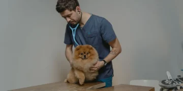










Quick take: An eye ulcer (corneal ulcer) in dogs is a painful break in the surface of the eye’s clear front layer. It can range from a tiny scratch to a deep lesion that threatens vision. Early signs include redness, squinting, and a white spot on the cornea. Prompt veterinary care—often involving medicated eye drops and protective bandages—usually leads to full healing within a few weeks.
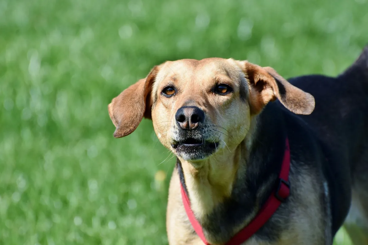
It’s 9 p.m., and your 7‑year‑old mixed‑breed dog, Milo, is lying on the couch with his head tilted to the side. He blinks slowly, avoids looking at the hallway, and you notice a faint white spot on his right eye. Your heart jumps. You’ve Googled “dog eye ulcer” and the pictures look scary. You wonder if this is something you can treat at home or if it’s an emergency.
We get it—eye problems feel urgent because you can see the discomfort right on your dog’s face. The good news is that most corneal ulcers respond well to treatment if caught early. In this guide we’ll explain what an eye ulcer is, how to spot it, what your vet will do, and how you can help your pup heal at home. We’ll also cover costs, breed risks, nutrition, and ways to prevent future ulcers.
What is eye ulcer in dogs?
An eye ulcer, technically a corneal ulcer, is a break in the cornea—the clear, dome‑shaped front window of the eye. The cornea protects the inner eye and helps focus light. When a tiny piece of tissue is lost, the underlying layers are exposed to air, bacteria, and tears, which can cause pain, infection, and scarring.
Corneal ulcers are fairly common in dogs. The American College of Veterinary Ophthalmology (ACVO) estimates that 10–15 % of dogs seen at general practices develop a corneal ulcer at some point in their lives. Small breeds such as Poodles, Cocker Spaniels, and Bulldogs are over‑represented, but any dog can develop an ulcer after trauma or disease.
What causes it?
Ulcers arise when the protective corneal surface is compromised. The main categories are:
| Cause | Typical examples |
|---|---|
| Physical trauma | Scratches from branches, fights with other dogs, or accidental pokes from toys |
| Infection | Bacterial (Staphylococcus, Pseudomonas), fungal (Candida), or viral (herpes‑like) agents |
| Dry eye (keratoconjunctivitis sicca) | Insufficient tear production, common in breeds like Shih Tzus and Bulldogs |
| Underlying eye disease | Entropion (inward‑rolling eyelid), corneal dystrophy, or chronic inflammation |
| Breed predisposition | Flat‑faced breeds, Poodles, and Cocker Spaniels often have structural eye issues that increase ulcer risk |
Signs and symptoms
Because the cornea is so sensitive, even a tiny ulcer can cause noticeable changes. Below is a quick guide to what you might see, from mild to severe.
| Severity | What you may notice |
|---|---|
| Mild | Redness (hyperemia), occasional squinting, a small white spot on the cornea, mild tearing |
| Moderate | More persistent squinting, obvious white or gray ulcer area, increased tear production, pawing at the eye, reluctance to play |
| Severe | Deep ulcer with a dark center, intense pain (dog keeps the eye shut), discharge that is thick or colored, corneal opacity, possible loss of vision |
When to call your vet
Call your vet today if you see any of the following:
- Redness or swelling of the eye
- Visible white or gray spot on the cornea
- Squinting, blinking, or pawing at the eye
- Excessive tearing or discharge
Go to an emergency veterinary hospital right now if you notice any of these red‑flag signs:
- The eye is completely shut or the dog refuses to open it
- There is a deep, dark ulcer with a large central defect
- Sudden loss of vision or the eye looks cloudy
- Severe pain indicated by vocalization, shaking, or inability to eat
This article is for information only and does not replace professional veterinary advice. Always consult your veterinarian for a proper diagnosis and treatment plan.
How vets diagnose it
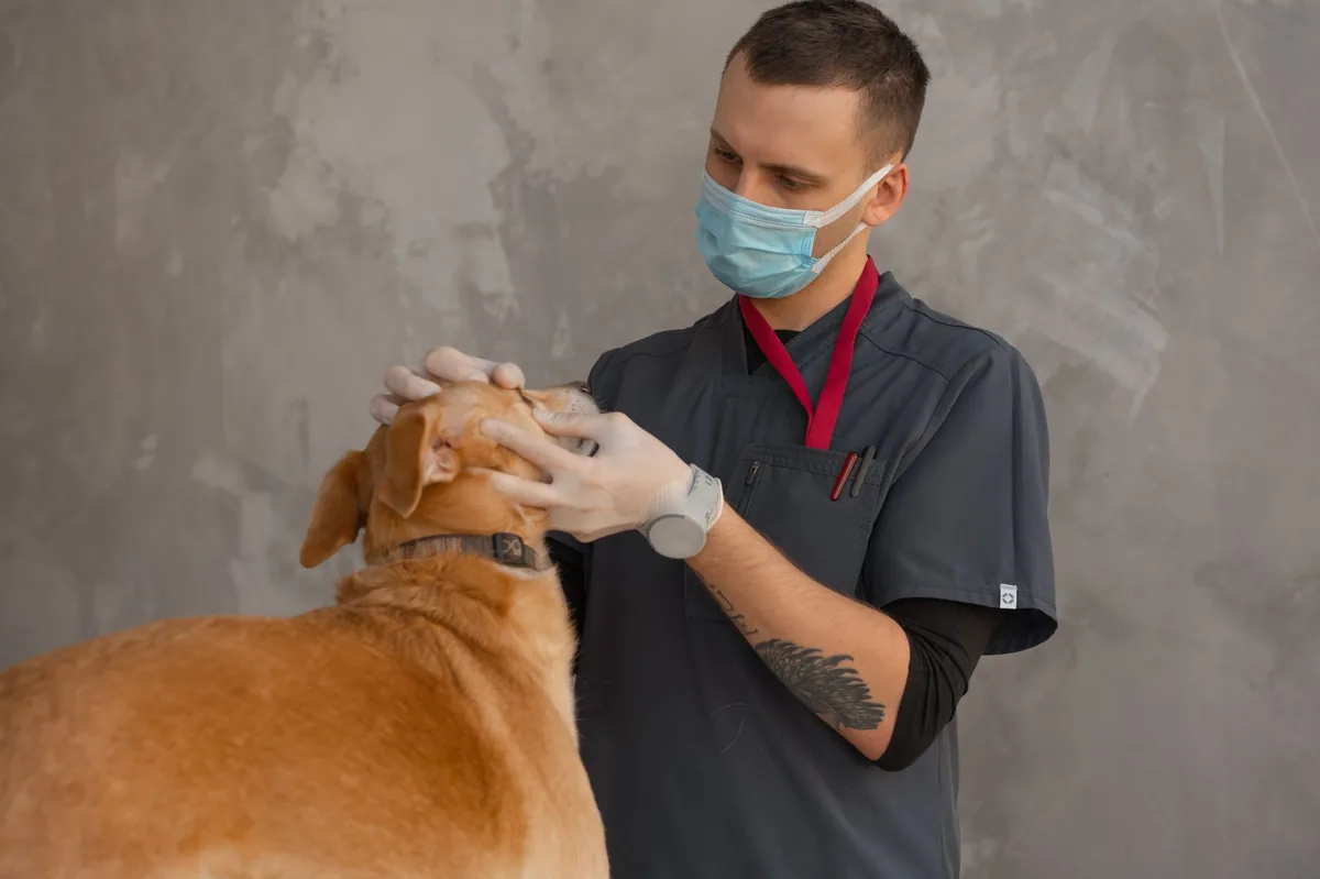
When you bring Milo in, the vet will start with a thorough history: when the signs began, any recent injuries, and your dog’s normal eye health. The physical exam includes a gentle eye exam using a slit‑lamp (a magnifying microscope) to look at the cornea’s surface.
The most common diagnostic test is fluorescein staining. A tiny amount of fluorescein dye is placed on the eye; the dye adheres to any gaps in the corneal epithelium, lighting up the ulcer under a blue light. This lets the vet grade the ulcer’s depth (grade I‑IV). If infection is suspected, a culture of the ulcer surface may be taken to identify bacteria or fungi, guiding antibiotic choice.
In complex cases, especially when vision is at risk, the vet might perform an ocular ultrasound or even a corneal topography (mapping the cornea’s shape) to assess underlying disease.
Treatment options
Medical treatment
Most ulcers heal with intensive medical therapy. The cornerstone is a regimen of medicated eye drops that protect the cornea, control infection, and reduce inflammation. Common drug classes include:
- Antibiotic drops such as moxifloxacin or tobramycin to prevent bacterial invasion.
- Antifungal drops (e.g., natamycin) if a fungal infection is confirmed.
- Anti‑inflammatory drops like prednisolone acetate, used cautiously and usually after the ulcer’s surface has begun to close.
- Lubricating ointments (e.g., artificial tears, ophthalmic gels) to keep the eye moist and promote epithelial growth.
For deeper ulcers, a protective bandage contact lens may be placed to shield the area while the medication works. Pain control is essential; oral NSAIDs (such as carprofen) or prescribed opioids can be used under veterinary guidance. Always ask your vet about each medication—dosage and frequency depend on your dog’s weight and ulcer grade.
Supplements and supportive care
While supplements can’t replace prescription meds, they may aid healing. Omega‑3 fatty acids (EPA/DHA) from fish oil have anti‑inflammatory properties and support corneal cell regeneration. A daily dose of a high‑quality fish‑oil supplement, as recommended by your vet, can be beneficial during recovery.
Probiotics are not directly linked to corneal healing, but maintaining overall gut health helps the immune system stay balanced, which can indirectly reduce the risk of secondary infections.
Procedures or surgery
When an ulcer is deep (grade III–IV), fails to respond to drops, or is caused by a structural problem, surgery may be needed. Options include:
- Corneal suturing (sutured keratoplasty) – stitching the cornea to close the defect.
- Superficial keratectomy – removing the damaged tissue and allowing a fresh surface to grow.
- Conjunctival flap – covering the ulcer with tissue from the inner eyelid to promote healing.
These procedures are performed under general anesthesia, and most dogs recover within 2–4 weeks. Post‑operative care involves the same eye drops and strict activity restriction.
Diet and nutrition
Healing a corneal ulcer isn’t just about eye drops; the body needs proper nutrients to rebuild tissue. While there is no “special ulcer diet,” feeding a balanced, high‑quality diet supports overall recovery.
Protein is the building block for new corneal cells. Look for foods that list a named animal protein (chicken, salmon, lamb) as the first ingredient, and aim for a minimum of 22 % protein on a dry‑matter basis. For dogs with dry eye or chronic inflammation, omega‑3 enriched formulas can be a good choice.
Antioxidants such as vitamin E and lutein help protect ocular cells from oxidative stress. Many premium kibble brands include added antioxidants; if yours does not, a supplement containing vitamin E (50–100 IU per day for a medium‑size dog) can be discussed with your vet.
Avoid foods high in sodium or artificial preservatives, as they can exacerbate inflammation. Also, limit treats that are rich in fat or sugar; excess calories can lead to weight gain, which may increase the risk of secondary eye problems like glaucoma.
Hydration is key. Encourage fresh water at all times, and consider adding a little low‑sodium broth to the bowl if your dog is a picky eater during recovery.
| Do feed | Limit | Avoid |
|---|---|---|
| High‑quality protein sources (chicken, fish, turkey) | Very fatty treats | Highly processed kibble with >10 % filler (corn, wheat) |
| Omega‑3 enriched foods or supplements | Excessive cheese or greasy foods | Artificial additives, excessive salt |
| Foods rich in antioxidants (blueberries, carrots) | Large bone meals (risk of eye trauma) | Any food that triggers allergies or GI upset |
When transitioning to a new or therapeutic diet, do it gradually over 5–7 days: mix 25 % new food with 75 % old food, then increase the new proportion every two days. This helps maintain gut health and keeps your dog comfortable while the eye heals.
Cost and prognosis
Eye‑ulcer treatment costs vary widely based on ulcer depth, need for surgery, and geographic location. In the United States, owners typically spend:
- Initial exam and diagnostics: $150–$300 (includes fluorescein staining, slit‑lamp exam, and possible culture).
- Medical management (drops, bandage lens, pain meds): $100–$250 for a two‑week course.
- Surgical intervention (if required): $800–$2,000, depending on the procedure and anesthesia fees.
In the United Kingdom, the same services range from £120–£250 for diagnostics, £80–£180 for medication, and £600–£1,500 for surgery. Many pet insurance plans cover a portion of these expenses, especially if the ulcer is classified as “acute injury.” Always check your policy’s exclusions (e.g., pre‑existing conditions).
Prognosis is excellent for superficial ulcers—most heal within 7–14 days with proper care. Deeper ulcers may take 3–4 weeks, and some can lead to permanent scarring that blurs vision. Early treatment dramatically improves outcomes; delayed care increases the risk of infection, perforation, and loss of the eye.
Prevention and home care
After Milo’s ulcer heals, you’ll want to keep his eyes safe. Here are everyday steps that reduce the chance of a repeat:
- Trim nails and keep toys smooth. Rough toys or sharp objects are common culprits for accidental scratches.
- Manage dry eye. If your vet diagnosed keratoconjunctivitis sicca, follow the prescribed tear‑substituting drops daily.
- Monitor breed‑specific risks. Flat‑faced breeds benefit from regular eyelid checks for entropion or lagophthalmos (incomplete eyelid closure).
- Protect eyes after surgery. Use an Elizabethan collar (e‑collar) and keep the head clean to avoid debris entering the wound.
- Schedule routine eye exams. A yearly check with your vet or a veterinary ophthalmologist catches early changes before they become ulcers.
During recovery, keep Milo’s environment calm. Limit vigorous play, especially jumping or running in dusty areas, for at least two weeks. Use a soft, breathable e‑collar to stop him from pawing at the eye, and apply the prescribed drops exactly as instructed.
From our vet team: “If you notice a white spot on the cornea, don’t wait for it to get bigger. Early intervention with fluorescein staining often reveals a tiny ulcer that can be closed with drops alone. Most owners are surprised at how quickly the eye can heal when the regimen is followed faithfully.”
Key takeaways
- Eye ulcers are breaks in the cornea that cause pain, redness, and a white spot; they require veterinary evaluation.
- Common causes include trauma, infection, dry eye, and breed‑related eyelid problems.
- Prompt treatment with medicated drops and protective lenses heals most superficial ulcers within 1–2 weeks.
- Deep or infected ulcers may need surgery; costs range from a few hundred to a few thousand dollars/pounds.
- Feed a high‑protein, antioxidant‑rich diet and consider omega‑3 supplements to support tissue repair.
- Prevent future ulcers by keeping toys smooth, managing dry eye, and scheduling regular eye exams.
Myth vs. fact
Myth: “All eye ulcers look the same as a simple pink eye.”
Fact: Conjunctivitis (pink eye) causes redness and discharge but does not produce a white corneal lesion. An ulcer will show a distinct white or gray spot on the cornea that stains with fluorescein.
Myth: “If the eye looks okay, the ulcer will heal on its own.”
Fact: Even a small ulcer can become infected or deepen quickly. Veterinary treatment accelerates healing and prevents permanent scarring.
Myth: “Eye drops are the only thing needed; diet doesn’t matter.”
Fact: Proper nutrition provides the building blocks for corneal cell regeneration; omega‑3 fatty acids and antioxidants can reduce inflammation and improve outcomes.
Frequently asked questions
What causes eye ulcers in dogs?
Eye ulcers result from any event that damages the corneal surface—scratches from branches, fights, foreign bodies, infections, dry eye, or underlying eyelid disorders. Breed anatomy can also predispose certain dogs to repeated trauma.
How can I tell if my dog has an eye ulcer?
The quickest clue is a white or gray spot on the clear part of the eye that stains bright blue with fluorescein dye. Accompanying signs include squinting, excessive tearing, and a red, inflamed eye. A vet’s exam is needed for confirmation.
Can an eye ulcer heal without veterinary care?
Very superficial scratches may close on their own, but most ulcers need medicated drops to prevent infection and scarring. Untreated ulcers can perforate, leading to vision loss or loss of the eye.
What is the typical recovery time for a dog’s eye ulcer?
Superficial (grade I–II) ulcers often heal in 7–14 days with drops. Deeper ulcers (grade III–IV) may require 3–4 weeks of treatment, and surgical cases can extend to 6 weeks for full recovery.
Are there any long‑term complications from a corneal ulcer in dogs?
If the ulcer heals without scarring, vision remains normal. However, deep ulcers can leave a scar that causes haze or astigmatism, and severe cases may lead to secondary glaucoma or loss of the eye.
How much does treatment for a dog eye ulcer usually cost?
In the U.S., basic diagnostics and medication typically run $250–$550, while surgery adds $800–$2,000. In the U.K., expect £200–£400 for non‑surgical care and £600–£1,500 for operative treatment. Check with your pet insurance for coverage details.
Ask the PuppaDogs community
Have a question this article didn’t fully answer? Want to compare notes with other dog owners who’ve been through this? Our community forum is moderated by experienced owners and vets — and answers tend to come fast. Ask in the PuppaDogs community →
References
- American College of Veterinary Ophthalmology (ACVO). “Corneal Ulcer Overview.” 2023.
- Merck Veterinary Manual. “Corneal Ulcer, Diagnosis and Treatment.” 2022.
- American Animal Hospital Association (AAHA). “Guidelines for Ocular Health.” 2021.
- Cornell University College of Veterinary Medicine. “Canine Dry Eye (KCS) and Corneal Ulcers.” 2022.
- World Small Animal Veterinary Association (WSAVA). “Ophthalmic Care Recommendations.” 2023.
- Veterinary Ophthalmology, 5th Edition. Gelatt, Knapp, and Grahn. 2020.
- American Veterinary Medical Association (AVMA). “Pet Insurance and Ocular Procedures.” 2022.


