













Quick take: Eye inflammation affecting the choroid and retina in dogs is a serious but treatable condition. Early signs include cloudy eyes, pupil changes, and vision loss. Prompt veterinary care—often involving eye exams, imaging, and targeted medication—greatly improves the chance of preserving sight.
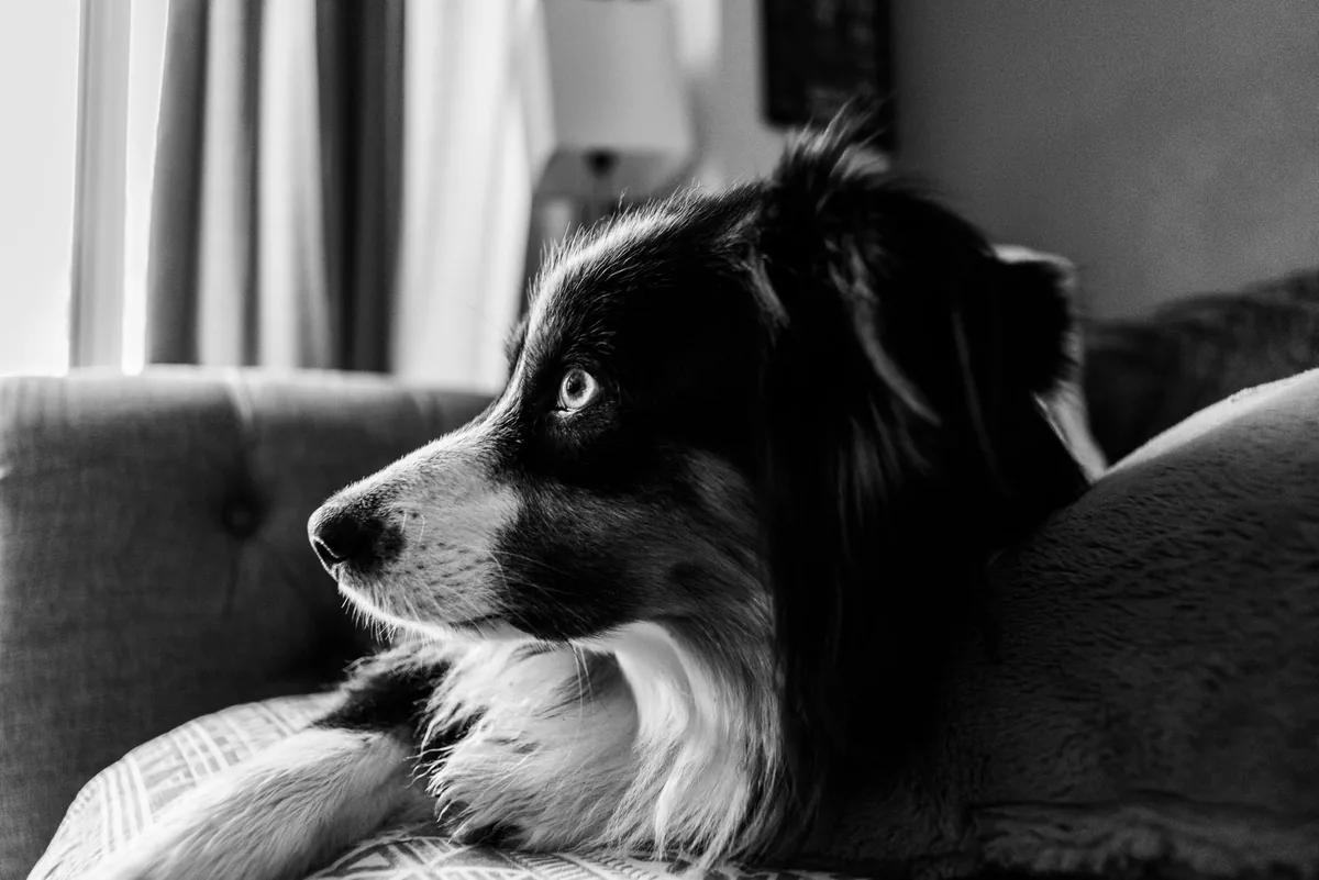
It’s 9 p.m., the lights are dimmed, and your usually energetic Border Collie, Max, is lying on the couch, staring blankly at the wall. You notice his left eye looks milky, and when you gently press his eyelid, a tiny ripple spreads across the surface. Your heart starts racing, and the Google search bar opens before you even finish the sentence “why is my dog’s eye…”.
First, take a breath. You’re not alone—many owners experience that exact moment of alarm. In most cases, choroiditis or retinal inflammation can be diagnosed and managed before permanent blindness sets in. Below we’ll walk through what this eye inflammation means, what to watch for, how vets figure it out, and the steps you can take to protect Max’s vision.
What is eye inflammation (choroid and retina) in dogs?
The choroid is a thin, vascular layer that sits behind the retina, supplying oxygen and nutrients to the light‑sensing cells. The retina itself is the inner lining of the eye that captures images and sends visual signals to the brain. When either of these layers becomes inflamed—often called choroiditis (choroid inflammation) or retinitis (retinal inflammation)—the normal flow of blood and nutrients is disrupted, leading to pain, cloudiness, and potentially permanent vision loss.
According to the American College of Veterinary Ophthalmologists (ACVO), choroidal and retinal inflammation together account for roughly 5–10 % of ocular disease cases seen in referral hospitals. While any breed can be affected, certain breeds such as the Siberian Husky, English Springer Spaniel, and German Shepherd are reported more often in peer‑reviewed case series.
What causes it?
Inflammation of the choroid and retina can stem from infectious agents, immune‑mediated disorders, or systemic diseases. Below is a concise breakdown:
| Category | Typical Causes | Notes for Owners |
|---|---|---|
| Infectious | Leptospira, Toxoplasma gondii, Neospora caninum, fungal organisms (Cryptococcus), bacterial infections (Streptococcus, Bartonella) | Often linked to exposure to standing water, raw diets, or tick bites. |
| Immune‑mediated | Autoimmune uveitis, systemic lupus erythematosus, idiopathic (unknown origin) choroiditis | May appear without any obvious trigger; blood tests can reveal underlying immune activity. |
| Systemic disease | Hypertension, diabetes mellitus, hyperlipidemia, neoplasia (cancers that spread to the eye) | Managing the underlying disease is essential for eye health. |
| Trauma | Blunt or penetrating eye injury | Even a minor scratch can set off inflammation if the protective layers are breached. |
Signs and symptoms
Recognizing early signs lets you act before the inflammation progresses. Dogs can’t tell us they’re in pain, so look for subtle changes in behavior and appearance.
| Severity | Typical Signs |
|---|---|
| Mild | Cloudy or slightly hazy appearance, occasional squinting, mild redness (conjunctival injection), reduced interest in toys. |
| Moderate | Noticeable pupil dilation or constriction, persistent squinting, visible “floaters” or “shadows” in vision, reluctance to climb stairs. |
| Severe | Marked opacity of the eye (white or yellow), severe pain when the eye is touched, sudden blindness, hemorrhage in the retina, excessive tearing. |
Other red‑flag clues include:
- Sudden loss of the “follow‑the‑hand” response.
- Visible blood vessels that look “corkscrew” or abnormal.
- Changes in eye color or a yellowish discharge.
- Behavioral shifts—becoming more timid, bumping into furniture, or hesitating to jump.
When to call your vet
Call your vet today if you notice any of the mild‑to‑moderate signs listed above—especially cloudiness, pupil changes, or a new reluctance to play.
Go to an emergency veterinary hospital right now if your dog shows severe pain, sudden blindness, a large amount of discharge, or visible bleeding inside the eye. These are emergencies that can lead to permanent vision loss within hours.
This article is for informational purposes only and does not replace a hands‑on examination by a licensed veterinarian.
How vets diagnose it
Diagnosing choroiditis or retinitis involves a stepwise approach that combines a thorough history with specialized tests.
- History & physical exam: Your vet will ask about recent illnesses, travel, tick exposure, diet changes, and any systemic symptoms like fever or weight loss.
- Complete ocular exam: Using an ophthalmoscope, the vet evaluates the cornea, pupil, lens, and the back of the eye (fundus). A fluorescein stain may highlight corneal ulcers.
- Ultrasound (B‑mode): This non‑invasive imaging sees the choroid and retina even when the eye is cloudy.
- Optical coherence tomography (OCT): Provides high‑resolution cross‑sections of retinal layers, useful for measuring thickness.
- Laboratory testing: Blood work (CBC, chemistry panel) can reveal infections or systemic disease. Specific serology (e.g., Leptospira MAT, Toxoplasma IgG/IgM) helps pinpoint infectious agents.
- Polymerase chain reaction (PCR) of ocular fluid: In select cases, a sample of aqueous humor is tested for bacterial or protozoal DNA.
Treatment options
Medical treatment
Most cases are managed medically. The goal is to reduce inflammation, treat any underlying infection, and protect the eye from secondary damage.
- Anti‑inflammatory drugs: Steroids such as prednisolone are the cornerstone for immune‑mediated inflammation. Your vet may also prescribe a non‑steroidal anti‑inflammatory drug (NSAID) like meloxicam for pain relief.
- Immunosuppressants: When steroids alone aren’t enough, drugs like ciclosporin or mycophenolate mofetil may be added. These help calm an overactive immune system.
- Antimicrobials: If an infection is identified, targeted antibiotics (e.g., doxycycline for Leptospira) or antiparasitics (e.g., clindamycin for Toxoplasma) are prescribed. Ask your vet about this option.
- Topical eye drops: Cyclosporine ophthalmic solution or a steroid eye drop can be used for localized control.
Supplements and supportive care
Adjunctive supplements can aid recovery, though they should never replace prescription medication.
- Omega‑3 fatty acids (EPA/DHA): Found in fish oil, they have anti‑inflammatory properties and may improve retinal health. A daily dose of 20–40 mg EPA per kilogram of body weight is common, but ask your vet for the right amount.
- Antioxidants: Vitamin E and lutein are sometimes recommended to protect retinal cells from oxidative stress.
- Probiotics: If systemic disease (e.g., diabetes) is a factor, a high‑quality probiotic can support overall immune balance.
Procedures or surgery
In rare, advanced cases, surgical intervention may be required.
- Vitrectomy: Removal of inflamed vitreous humor to clear the visual axis. Recovery can take 2–4 weeks and costs typically range from $2,500–$4,500 (U.S.) or £2,000–£3,500 (U.K.).
- Retinal detachment repair: If the retina separates due to severe inflammation, a retinal re‑attachment surgery may be performed. Success rates vary, and the procedure is expensive (often $3,000–$6,000).
Diet and nutrition
While no single “eye‑cure” diet exists, certain nutritional strategies can support retinal health and reduce inflammation.
Key nutrients for retinal support
- Highly digestible protein: Ensures the body has the building blocks needed for tissue repair without overloading the kidneys. Choose a commercial diet that lists a named meat source first.
- Omega‑3 fatty acids: As mentioned, EPA and DHA help modulate inflammation. Look for dog foods that contain fish oil or algal oil as a primary ingredient.
- Antioxidants: Vitamins C and E, beta‑carotene, lutein, and zeaxanthin protect photoreceptor cells from oxidative damage. Some therapeutic diets (e.g., “ophthalmic” formulas) fortify these compounds.
- Low‑sodium and low‑carbohydrate: Helps control systemic hypertension and diabetes—two common contributors to retinal disease.
Foods to favor, limit, or avoid
| Do feed | Limit | Avoid |
|---|---|---|
| High‑quality kibble or wet food with real meat, fish, or poultry as the first ingredient. | Table scraps high in fat or salt (e.g., bacon, cheese). | Raw bones that could cause ocular trauma. |
| Supplemental fish oil (approved for dogs) or canned fish in water. | Excessive treats, especially those high in sugar. | Artificial food colorings and preservatives. |
| Fresh vegetables like carrots and leafy greens for added antioxidants. | High‑glycemic carbs (white rice, corn). | Commercial “grain‑free” diets that lack scientific backing for eye health. |
Feeding tips during recovery
- Offer smaller, more frequent meals to reduce the workload on the digestive system.
- Warm the food slightly (to about body temperature) to enhance aroma, encouraging a dog with reduced appetite to eat.
- Transition to any new therapeutic diet gradually over 7–10 days—mixing 25 % new food with 75 % old, then increasing the new portion each few days.
- Ensure fresh water is always available; hydration supports overall healing.

For dogs with severe systemic disease, your veterinarian may recommend a prescription “renal” or “diabetic” diet that is low in phosphorus and simple sugars. These diets are formulated to reduce stress on the eye’s blood supply while still providing balanced nutrition.
Cost and prognosis
Financial considerations are an important part of planning care. Below is a rough breakdown of typical expenses (all figures are estimates and can vary by region and clinic).
| Service | U.S. Estimate | U.K. Estimate |
|---|---|---|
| Initial ophthalmic exam (including dilated fundus exam) | $150–$300 | £120–£250 |
| Ultrasound or OCT imaging | $200–$500 | £180–£400 |
| Blood work & serology | $100–$250 | £80–£200 |
| Medical therapy (steroids, immunosuppressants, antibiotics – 4‑week course) | $100–$400 | £80–£350 |
| Surgical intervention (vitrectomy or retinal repair) | $2,500–$6,000 | £2,000–£5,000 |
Pet insurance often covers a portion of the diagnostic work‑up and medication, especially if the policy includes “eye care” or “specialist referral” clauses. When obtaining a quote, ask the insurer whether pre‑existing conditions are excluded and if there’s a waiting period for ophthalmic claims.
Prognosis depends on the underlying cause and how quickly treatment starts. Immune‑mediated choroiditis caught early has a 70–80 % chance of preserving vision, while infectious forms that progress to retinal detachment carry a poorer outlook. Most dogs that respond to therapy regain functional vision within 4–8 weeks, though some may retain a slight haze or reduced night vision.
Prevention and home care
While not all cases are preventable, many risk factors can be mitigated with everyday vigilance.
- Tick control: Use veterinarian‑recommended products year‑round, especially in high‑risk regions. (See our Dog Questions Answered guide for tick‑prevention options.)
- Vaccination: Keep core vaccines up to date; leptospirosis vaccines reduce the risk of Leptospira‑related eye disease.
- Regular veterinary exams: Annual eye checks for senior dogs can catch early inflammation before it becomes symptomatic.
- Balanced diet: Feeding a diet rich in omega‑3s and antioxidants supports retinal health.
- Safe environment: Prevent eye trauma by keeping sharp objects out of reach and supervising play with other dogs.
At home, monitor your dog’s vision by gently moving a favorite toy across the room and noting whether he tracks it. Keep a daily log of any changes in eye appearance, behavior, or appetite, and share this with your vet at follow‑up visits.
From our vet team: Early detection is the biggest game‑changer. If you notice even a subtle cloudiness or a change in your dog’s response to visual cues, schedule a vet appointment right away. Most inflammation can be halted before it damages the retina, and the sooner we start treatment, the better the odds of keeping your companion’s world bright.
Key takeaways
- Eye inflammation of the choroid or retina is serious but often treatable if caught early.
- Watch for cloudiness, pupil changes, and any sudden loss of visual response.
- Immediate veterinary evaluation—especially for severe pain or blindness—can prevent permanent vision loss.
- Treatment typically involves steroids, targeted antimicrobials, and supportive supplements like fish oil.
- A diet rich in high‑quality protein, omega‑3 fatty acids, and antioxidants helps the healing process.
- Regular eye exams, tick control, and vaccination are key preventive measures.
Myth vs. fact
Myth: All eye inflammation in dogs leads to blindness.
Fact: While untreated inflammation can cause blindness, many cases respond well to medication, especially when diagnosed early.
Myth: Only older dogs get choroiditis.
Fact: Both young and senior dogs can develop inflammation; infections or immune triggers can affect any age.
Myth: Eye drops alone cure the condition.
Fact: Systemic medications (steroids, immunosuppressants) are usually required, and eye drops are used as an adjunct.
Frequently asked questions
What causes choroid and retina inflammation in dogs?
It can be triggered by infections (like Leptospira, Toxoplasma), immune‑mediated diseases, systemic illnesses such as hypertension, or direct trauma to the eye.
What symptoms should I look for if my dog has eye inflammation?
Look for cloudy eyes, pupil dilation or constriction, excessive tearing, squinting, and any sudden change in visual behavior such as bumping into objects.
Can retinal inflammation lead to blindness in dogs?
Yes, if the inflammation is severe or left untreated it can damage the retina permanently, resulting in partial or total blindness. Prompt treatment greatly reduces this risk.
How is eye inflammation diagnosed by a veterinarian?
The vet will perform a dilated fundus exam, possibly use ultrasound or OCT imaging, and run blood work or ocular fluid PCR to identify infectious agents or systemic disease.
What are the treatment options for choroiditis in dogs?
Standard care includes systemic steroids, immunosuppressants, and targeted antibiotics or antiparasitics. Supportive supplements like omega‑3 fish oil are often added, and surgery is reserved for advanced cases.
Is there a way to prevent eye inflammation in at‑risk breeds?
Regular eye exams, diligent tick control, staying current on core vaccinations, and feeding a diet rich in antioxidants can lower the likelihood of inflammation, especially in predisposed breeds like Siberian Huskies and English Springer Spaniels.
Ask the PuppaDogs community
Have a question this article didn’t fully answer? Want to compare notes with other dog owners who’ve been through this? Our community forum is moderated by experienced owners and vets — and answers tend to come fast. Ask in the PuppaDogs community →
References
- American College of Veterinary Ophthalmologists (ACVO). “Guidelines for the Diagnosis and Treatment of Inflammatory Eye Disease in Dogs.” 2023.
- American Animal Hospital Association (AAHA). “Canine Ophthalmology Standards.” 2022.
- Merck Veterinary Manual. “Choroiditis and Retinitis in Dogs.” Updated 2023.
- World Small Animal Veterinary Association (WSAVA). “Nutritional Recommendations for Dogs with Ocular Disease.” 2021.
- American Veterinary Medical Association (AVMA). “Leptospirosis Vaccination and Ocular Manifestations.” 2022.
- University of California, Davis School of Veterinary Medicine. “Ocular Imaging Techniques in Small Animals.” 2022.
- American College of Veterinary Internal Medicine (ACVIM). “Systemic Hypertension in Dogs: Implications for Eye Health.” 2023.
- Plumb’s Veterinary Drug Handbook. “Steroids and Immunosuppressants for Ophthalmic Use.” 9th Edition, 2021.
- American Kennel Club (AKC). “Breed Predispositions to Ocular Inflammation.” 2022.
- Pet Insurance Review. “Coverage for Ophthalmic Procedures.” 2023.


