











Quick take: Pannus is a progressive, immune‑mediated inflammation of the cornea that shows up as a gray‑white, vascularized haze on the front of the eye. It’s most common in older, light‑colored dogs and can lead to vision loss if untreated. Early detection, regular vet checks, and a combination of eye drops, supplements, and sometimes surgery keep most dogs comfortable and preserve sight.
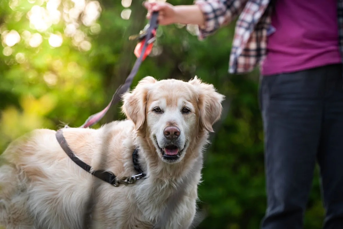
It’s 9 p.m., the house is quiet, and your senior Golden Retriever, Max, is lying on the couch while you’re scrolling through search results. You notice a faint, cloudy spot on his right eye and a slight squint when you gently touch his eyelid. Your heart races—“Is this something serious?” you wonder. You’re not alone; many owners first spot pannus during a routine grooming or a late‑night cuddle.
We understand that a hazy cornea can feel alarming, especially when you hear words like “vascularization” or “immune‑mediated.” The good news is that pannus is usually manageable, especially when caught early. In this article we’ll explain exactly what pannus is, why it shows up in certain breeds, how to tell the difference from other eye problems, what your vet will do to diagnose it, and the full spectrum of medical, supplemental, and surgical options—including costs and long‑term care.
By the end you’ll know the signs to watch for, when to call the vet, how diet and lifestyle can support recovery, and what realistic outlook to expect for your furry companion.
What is pannus in dogs?
Pannus (also called “corneal vascularization” or “chronic superficial keratitis”) is a progressive inflammation of the cornea—the clear, outer layer of the eye. In pannus, immune cells trigger new blood vessels to grow into the normally avascular cornea, creating a gray‑white, “frosted” appearance. The new vessels bring inflammatory cells that can scar the cornea and, over time, impair vision.
While pannus can occur at any age, it most often affects senior dogs (usually 7 years and older) with light‑colored coats and eyes. The American College of Veterinary Ophthalmologists (ACVO) notes that pannus accounts for roughly 5‑10 % of ophthalmic cases seen in referral hospitals, making it a relatively common, though not universal, problem.
What causes it?
- Immune‑mediated disease: The primary driver is an abnormal immune response that mistakenly attacks the corneal tissue.
- Genetic predisposition: Certain breeds—especially those with light eyes—have a higher inherited risk. See the table below for the most affected breeds.
- Chronic ocular irritation: Long‑standing dry eye (keratoconjunctivitis sicca), allergies, or exposure to UV light can exacerbate inflammation.
- Environmental factors: High UV exposure, dusty or windy environments, and persistent irritants (e.g., smoke) can worsen the condition.
| Breed (high risk) | Typical age of onset |
|---|---|
| Old English Sheepdog | 7‑9 years |
| Collie (including Border Collie) | 6‑8 years |
| Greyhound | 7‑10 years |
| Akita | 8‑10 years |
| Australian Shepherd | 6‑9 years |
Signs and symptoms
Early pannus can be subtle, but the disease progresses through recognizable stages. Spotting the first changes lets you seek treatment before scarring develops.
| Stage | What you’ll see |
|---|---|
| Early (mild) | Fine, gray‑white haze near the edges of the cornea; occasional squinting. |
| Moderate | More extensive haze covering the central cornea; visible tiny blood vessels (punctate vessels); increased tearing. |
| Severe | Dense, opaque cloudiness, obvious vascular growth, possible ulceration, and noticeable vision loss. |
Other clues include:
- Frequent blinking or rubbing the eye with the paw.
- Redness of the surrounding conjunctiva (the pink part of the eye).
- Discomfort when bright lights are on.
- Changes in behavior—reluctance to play fetch or navigate stairs.
When to call your vet
Call your vet today (non‑emergency) if you notice any of the following:
- Persistent gray‑white haze or any new blood vessels on the cornea.
- Frequent tearing, squinting, or rubbing of the eye.
- Signs of discomfort such as whimpering when the eye is touched.
Go to an emergency veterinary hospital right now if you see:
- Sudden loss of vision or a dog stumbling into furniture.
- Corneal ulceration (a raw, painful spot) or a sudden increase in redness.
- Excessive discharge that is thick, yellow, or foul‑smelling.
These red‑flag symptoms can indicate a complication that needs urgent care to preserve sight.
This article provides general information and is not a substitute for professional veterinary advice.
How vets diagnose it
Diagnosing pannus involves a systematic eye exam and a few ancillary tests:
- History and visual inspection: Your vet will ask when you first noticed the haze and will examine both eyes with a bright otoscope or slit lamp.
- Fluorescein staining: A drop of fluorescent dye highlights any corneal ulcers or erosions that can accompany pannus.
- Schirmer tear test: Measures tear production to rule out keratoconjunctivitis sicca (dry eye), which can look similar.
- Intra‑ocular pressure (tonometry): Ensures glaucoma is not present, as increased pressure can also cause cloudiness.
- Blood work (CBC, chemistry): Checks for systemic immune disorders or underlying infections that might trigger pannus.
All of these steps help the veterinarian differentiate pannus from other corneal diseases such as dry eye, ulceration, or fungal infections.
Treatment options
Medical treatment
The mainstay of therapy is topical anti‑inflammatory eye drops. The most commonly prescribed classes include:
- Corticosteroid drops: Prednisolone acetate or dexamethasone reduce inflammation quickly. Ask your vet about this option.
- Cyclosporine (Immunomodulator): Helps control immune‑mediated inflammation without the cataract‑risk associated with long‑term steroids. Ask your vet about this option.
- Non‑steroidal anti‑inflammatory drug (NSAID) drops: Such as flurbiprofen, useful for milder cases or as steroid‑sparing agents.
- Lubricating/artificial tears: Keep the cornea moist and aid healing, especially if dry eye is also present.
Most dogs need eye drops twice daily at first, tapering to once daily once the haze stabilizes. Your vet will schedule follow‑up exams every 2‑4 weeks during the active phase.
Supplements and supportive care
While supplements cannot replace prescription medication, they can bolster ocular health:
- Omega‑3 fatty acids (EPA/DHA): Found in fish‑oil capsules, they have anti‑inflammatory properties that may lessen corneal inflammation.
- Vitamin C and E: Antioxidants that support overall eye health; however, their effect on pannus is modest.
- Probiotics: Maintaining gut health can indirectly help immune regulation, especially in dogs with concurrent allergies.
Discuss any supplement with your vet to ensure appropriate dosing and to avoid interactions with prescribed eye drops.
Procedures or surgery
When medical therapy cannot halt progression, surgical options are considered:
- Superficial keratectomy (SK): Removes the affected outer corneal layer, allowing healthier tissue to regrow. Recovery is 2‑3 weeks, and the cost in the U.S. typically ranges from $800‑$1,500.
- Conjunctival graft or limbal stem‑cell transplant: Replaces damaged tissue with healthy tissue from the conjunctiva. This is more specialized and may cost $2,000‑$4,500.
- Corneal transplant (penetrating keratoplasty): Rarely performed for pannus, reserved for end‑stage disease with severe scarring. Costs can exceed $5,000 and require intensive post‑op care.
Most owners opt for medical management first; surgery is reserved for cases where vision is threatened despite aggressive eye‑drop therapy.
Diet and nutrition
Although pannus is primarily an immune‑mediated eye disease, nutrition influences overall inflammation and immune balance. Feeding a diet that supports a healthy immune system can complement medical therapy and may slow disease progression.
Here are practical feeding guidelines for dogs diagnosed with pannus:
| Do feed | Limit | Avoid |
|---|---|---|
| High‑quality protein from animal sources (chicken, turkey, fish). | Excessive treats high in salt. | Highly processed kibble with artificial colors and flavors. |
| Omega‑3 rich foods (salmon, sardines) or a vetted fish‑oil supplement. | Large amounts of omega‑6 fatty acids (corn oil, wheat germ). | Raw diets high in unsanitary meat unless carefully balanced. |
| Fresh vegetables low in oxalates (green beans, carrots) for antioxidants. | Excessive dairy (can aggravate inflammation in some dogs). | Grains with high gluten content if your dog shows a sensitivity. |
Key nutritional principles:
- Balanced, complete diet: Follow AAFCO standards to ensure all essential nutrients are present. Commercially prepared “premium” or “holistic” foods often meet these criteria.
- Moderate calories: Maintaining a healthy weight reduces systemic inflammation. Overweight dogs can have higher circulating inflammatory cytokines that may worsen pannus.
- Hydration: Adequate water intake supports tear production and overall ocular surface health.
- Prescription therapeutic diets: While no diet is specifically labeled for pannus, a “senior” or “anti‑inflammatory” formula from a reputable brand can be beneficial. Discuss options with your vet; they may recommend a renal‑support or joint‑support diet if your dog has concurrent issues.
Transition any new food over 7‑10 days, mixing increasing amounts of the new diet with decreasing amounts of the old one. This helps avoid gastrointestinal upset, which can stress the immune system further.
Cost and prognosis
| Item | U.S. estimate | U.K. estimate |
|---|---|---|
| Initial ophthalmic exam & basic diagnostics | $150‑$250 | £80‑£150 |
| Topical corticosteroid drops (30 days) | $30‑$70 | £20‑£50 |
| Cyclosporine eye drops (30 days) | $80‑$130 | £60‑£110 |
| Superficial keratectomy surgery | $800‑$1,500 | £600‑£1,200 |
| Follow‑up visits (each) | $70‑$120 | £40‑£90 |
Overall, most owners spend $300‑$800 in the first year for medication, exams, and any needed procedures. Surgical cases can push the total above $2,000.
Prognosis is generally good when pannus is caught early and managed with consistent eye drops. Studies referenced by the Merck Veterinary Manual indicate that 70‑80 % of dogs retain functional vision with proper treatment. However, if the disease progresses to severe scarring, permanent vision loss can occur.
Long‑term monitoring (every 6‑12 months after stabilization) helps catch relapses early. Many dogs live comfortably for years with pannus, especially when owners adhere to the treatment plan.
Prevention and home care
While you can’t completely prevent an immune‑mediated condition, several practical steps reduce the risk or slow progression:
- Regular veterinary eye exams: At least once a year for senior dogs, and more often if pannus has been diagnosed.
- UV protection: Limit prolonged exposure to bright sunlight; consider canine sunglasses for dogs that tolerate them.
- Control dry eye: If your dog has keratoconjunctivitis sicca, strict compliance with artificial tear therapy reduces secondary inflammation.
- Allergy management: Identify and avoid environmental allergens (pollens, dust) that can trigger ocular irritation.
- Maintain a healthy weight: Obesity fuels systemic inflammation, which can exacerbate pannus.
- Promptly address any eye trauma: Even a minor scratch can ignite an inflammatory cascade.
For owners who love outdoor adventures, a quick rinse of the eyes with sterile saline after a walk in dusty conditions can help keep the cornea clear.
From our vet team: “If your dog’s eyes look cloudy, don’t wait for the haze to spread. Early intervention with anti‑inflammatory drops often preserves vision, and most dogs respond well. Keep a small bottle of sterile saline at home, and schedule that first eye check within a week of noticing any change.”
Key takeaways
- Pannus is an immune‑mediated corneal inflammation that appears as a gray‑white haze and can lead to vision loss if untreated.
- Light‑colored, senior breeds such as Old English Sheepdogs and Collies are most at risk.
- Early signs include subtle haze, increased tearing, and squinting; seek veterinary care promptly.
- Medical management with corticosteroid or cyclosporine eye drops is the first‑line treatment; surgery is reserved for refractory cases.
- Supportive nutrition—high‑quality protein, omega‑3s, and antioxidants—helps control systemic inflammation.
- Regular eye exams, UV protection, and weight management are practical ways to prevent progression.
Myth vs. fact
Myth: Pannus always leads to blindness.
Fact: When caught early and treated consistently, most dogs retain functional vision; blindness is rare and usually linked to severe, untreated cases.
Myth: Only “old” dogs get pannus.
Fact: Although it’s most common in seniors, younger dogs with strong genetic predisposition can develop pannus, especially in breeds with light eyes.
Myth: Over‑the‑counter eye drops can cure pannus.
Fact: Prescription anti‑inflammatory drops are needed; OTC lubricants may help comfort but do not address the underlying immune process.
Frequently asked questions
What does pannus look like on a dog’s eye?
It appears as a gray‑white, cloudy haze that may start at the periphery and spread inward, often with tiny, fine blood vessels growing into the cornea.
Can pannus cause blindness in dogs?
Yes, but usually only if the disease progresses unchecked and leads to dense scarring; early treatment dramatically reduces this risk.
What causes pannus in certain breeds?
Genetic factors give light‑eyed breeds a higher tendency for an abnormal immune response that targets the cornea, combined with age‑related changes in ocular immunity.
How is pannus diagnosed by a veterinarian?
The vet performs a detailed eye exam with a slit lamp, uses fluorescein staining to rule out ulcers, measures tear production, checks intra‑ocular pressure, and may run blood work to look for systemic immune issues.
What are the most effective treatments for canine pannus?
Topical corticosteroid or cyclosporine eye drops are first‑line; if they fail, surgical options like superficial keratectomy are considered. Supplements such as omega‑3s can aid inflammation control.
How much does pannus treatment typically cost?
Initial diagnostics and medication usually run $300‑$800 in the U.S.; surgical interventions can raise total costs to $2,000‑$5,000 depending on the procedure and region.
Ask the PuppaDogs community
Have a question this article didn’t fully answer? Want to compare notes with other dog owners who’ve been through this? Our community forum is moderated by experienced owners and vets — and answers tend to come fast. Ask in the PuppaDogs community →
References
- American College of Veterinary Ophthalmologists (ACVO) – “Corneal Diseases in Dogs,” clinical guidelines.
- Merck Veterinary Manual – “Pannus (Chronic Superficial Keratitis) in Dogs” chapter.
- American Animal Hospital Association (AAHA) – “Ophthalmology Guidelines for Companion Animals,” 2023.
- World Small Animal Veterinary Association (WSAVA) – “Standard of Care for Canine Ophthalmic Diseases.”
- Veterinary Ophthalmology (3rd ed.) – Gelatt, 2022, sections on immune‑mediated corneal disease.
- American Veterinary Medical Association (AVMA) – “Dry Eye (Keratoconjunctivitis Sicca) in Dogs” fact sheet.
- University of California, Davis School of Veterinary Medicine – “Canine Ocular Immunology” research overview.
- AAFA (American Association of Feline Practitioners) – “Omega‑3 Fatty Acids and Inflammation,” adapted for canine use.


