


⚡ Quick answer: Dog mast cell tumour (MCT) pre-test probability calculator. Scores lump features (location, size, growth, fluctuation, Darrier’s sign), systemic signs and breed to indicate when fine-needle aspirate cytology is warranted – the most common malignant canine skin tumour.
Why Every New Lump Needs A Needle
Mast cell tumour (MCT) is the most common malignant skin tumour in dogs — about 16-21% of all cutaneous neoplasia. The defining problem with MCT: visual diagnosis is unreliable. Some MCTs look exactly like lipomas. Some “obvious” lipomas turn out to be MCTs. Some textbook-looking MCTs are benign histiocytomas.
The modern standard is “aspirate first, decide treatment second” — a fine-needle aspirate of any new persistent cutaneous mass costs little, takes minutes, requires no anaesthesia, and produces a diagnosis (or close to one) within 24-48 hours. Mast cells with their characteristic purple cytoplasmic granules are diagnostic on cytology.
This calculator scores pre-test probability of MCT from features owners can identify, to prioritise vet visits and emphasise when aspirate is especially urgent.
The Classic MCT Features
MCT is the great mimic — it can look like almost anything cutaneous. But certain features raise suspicion significantly:
Darrier’s Sign
The mass swells when handled or rubbed. Caused by histamine release from degranulating mast cells. Highly suggestive of MCT — few other lumps do this. The dog may also develop redness or wheal formation around the mass.
Fluctuating Size
The mass changes size from week to week — sometimes bigger, sometimes smaller. Again caused by intermittent degranulation. Stable-size masses are less typical of MCT; fluctuation strongly raises suspicion.
Location
Some locations carry traditionally higher-grade behaviour:
| Location | Behaviour |
|---|---|
| Perianal / scrotum / vulva | Higher-grade tendency |
| Oral / mucocutaneous junctions | Higher-grade tendency |
| Inguinal / preputial | Moderately elevated |
| Distal limb / paw | Average |
| Trunk / head / neck | Most common location, average grade |
Size
- <2 cm — most often well-differentiated
- 2-5 cm — intermediate
- >5 cm — more often higher-grade
Growth Rate
- Slow — more often low-grade
- Moderate — variable
- Rapid (visible change in days) — concerning for higher-grade
Systemic / Paraneoplastic Signs
Histamine and heparin release from degranulating mast cells can cause:
- Vomiting — gastric ulceration from histamine
- Melaena (black tarry stool) — GI bleeding
- Easy bruising — heparin release
- Reduced appetite
- Rarely: anaphylactic-like reactions when masses are manipulated
Breed Predispositions
The breeds most over-represented in canine MCT populations:
- Boxer — the standout (relative risk 4-8× average). Good news: Boxer MCTs tend to be lower-grade and more often surgically curable than average.
- Boston Terrier
- English Bulldog / French Bulldog
- Pug
- Golden Retriever
- Labrador Retriever
- Beagle
- Weimaraner
- Staffordshire Bull Terrier / Pit Bull
- Shar Pei — historically poor prognosis in this breed; modern grading may identify lower-risk cases
- Cocker Spaniel
- Miniature Schnauzer
For high-risk breeds (especially Boxers), monthly owner self-exam is appropriate — feel for any new lump and aspirate within 1-2 weeks of detection.
Diagnostic Workup
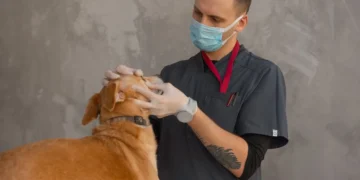
Step 1: Fine-Needle Aspirate Cytology
The definitive diagnostic test for MCT. Cheap, quick (often same-day result), no anaesthesia required:
- Fine needle into the mass with a syringe; gentle “wiggle” to collect cells
- Smear onto slide, stain with Diff-Quik or Wright’s
- Mast cells with purple cytoplasmic granules are diagnostic
- Usually 90%+ diagnostic for cutaneous MCT
A positive cytology confirms MCT but doesn’t grade it — that requires histopathology of the removed tumour.
Step 2: Surgical Excision With Wide Margins
The cornerstone treatment:
- 2-3 cm lateral margins around the visible mass
- One fascial plane deep
- Complete histopathological margins (“clean margins”) strongly predict local cure
- For tumours at extremities where margins are difficult, specialist surgical input is worthwhile
Step 3: Histopathology + Grading
Two grading systems used:
Patnaik 3-tier (historical, still used):
- Grade I — well-differentiated; usually surgical cure
- Grade II — intermediate; prognosis depends on mitotic index, KI67, location
- Grade III — poorly-differentiated; aggressive systemic disease likely
Kiupel 2-tier (newer, better inter-observer agreement):
- Low-grade — usually surgical cure
- High-grade — aggressive; needs adjuvant therapy
Most pathology reports now provide both grades.
Step 4: Staging (For Grade II+ Or High-Grade)
- Regional lymph node aspirate — the peripheral LN closest to the mass; detects nodal spread
- Abdominal ultrasound — spleen, liver, visceral lymph nodes (the typical metastatic sites)
- Buffy coat smear — historically used; poor sensitivity, less used now
- Chest radiographs — rare lung metastasis but for completeness
- c-KIT mutation testing — PCR or IHC for KIT pattern; mutated c-KIT predicts response to tyrosine kinase inhibitors
Treatment Options
Surgery Alone (Low-Grade MCT With Clean Margins)
~90% cure rate for low-grade MCT (Kiupel low / Patnaik I) with clean histopathological margins. No further treatment needed. Monitor for new MCTs at other sites (some dogs get multiple over a lifetime).
Surgery + Adjuvant Radiotherapy
For incomplete margins in dogs where wider re-excision is not feasible (e.g. distal limb). Excellent local control rate.
Surgery + Vinblastine + Prednisolone Chemotherapy
For high-grade or metastatic MCT. Vinblastine 2 mg/m² IV weekly x 4-8 weeks. Prednisolone tapered over 3-6 months. Reasonably well-tolerated; meaningful prolongation of survival in high-grade disease.
Tyrosine Kinase Inhibitors
For dogs with c-KIT mutations:
- Toceranib (Palladia) — daily oral; targets KIT, VEGFR, PDGFR
- Masitinib (Masivet) — daily oral; KIT-selective
Used for advanced or non-resectable MCT, or as adjuvant in high-grade disease. Excellent biologic activity in c-KIT-mutated tumours.
Palliative Care
For dogs where definitive treatment is not chosen — combination of prednisolone (often produces substantial palliative response), H1 + H2 blockers for paraneoplastic signs, pain management, supportive care.
Premedicate Before Manipulation
Because of degranulation risk, premedication is standard before any major manipulation of an MCT (FNA, biopsy, surgery):
- H1 blocker: diphenhydramine 2-4 mg/kg IM or chlorpheniramine
- H2 blocker: famotidine 1 mg/kg
- Steroid: prednisolone (sometimes dexamethasone IV preoperatively)
These reduce the risk of anaphylactic-like reactions and intraoperative hypotension.
Differentials For Cutaneous Lumps
What else could a lump be? Common differentials in adult dogs:
| Lump type | Features |
|---|---|
| Lipoma | Soft, mobile, slowly growing, typically older dogs, trunk |
| Sebaceous cyst | Often head/back; firm; may rupture with white/yellow discharge |
| Histiocytoma | Young dogs (<3 yrs); small red button on head/ears/limbs; often regresses spontaneously |
| Papilloma (wart) | Cauliflower-like; oral in young dogs, skin in adults |
| Sebaceous adenoma | Older dogs, head/trunk; small, may bleed when traumatised |
| Hair follicle tumour | Variable |
| Soft tissue sarcoma | Older dogs; firm; locally invasive; often limb |
| Squamous cell carcinoma | Older dogs; sun-exposed areas; ulcerated |
| Melanoma | Pigmented; oral or digital sites more aggressive |
| Histiocytic sarcoma | Aggressive; Bernese Mountain Dog and Rottweiler over-represented |
Aspirate distinguishes them all — usually within minutes.
Honest Caveats
- Visual diagnosis of cutaneous lumps is unreliable — aspirate every new persistent mass.
- Pre-test probability is not diagnosis — cytology is.
- A negative aspirate doesn’t always rule out MCT in atypical locations or low-cellularity samples — submit suspicious samples to a pathologist.
- Patnaik II MCT prognosis is variable — within this group, mitotic index, KI67, and c-KIT pattern subdivide further. The Kiupel 2-tier system handles this better.
- MCTs can be multiple — some dogs develop multiple MCTs over years. Each new lump still needs aspirate.
Conclusion
Mast cell tumour is the most common malignant canine skin cancer — and completely treatable in the majority of cases when caught early and managed properly. The defining features (Darrier’s sign, fluctuating size, paraneoplastic GI signs) raise pre-test probability, but the diagnosis is always made by fine-needle aspirate cytology. For at-risk breeds (Boxer, Bulldog, Pug, Golden Retriever, Labrador), monthly owner self-exam catches new lumps early when surgical cure is most likely. Aspirate every new persistent lump — the test costs little and changes treatment substantially when the answer is unexpected.
Frequently Asked Questions
What is Darrier’s sign in dogs?
Darrier’s sign is when a cutaneous mass swells, gets red, or develops wheal-like changes when handled, rubbed, or palpated. It’s caused by mast cell degranulation releasing histamine into surrounding tissue. Highly suggestive of MAST CELL TUMOUR (MCT) – few other cutaneous masses do this. Any lump with Darrier’s sign needs fine-needle aspirate cytology promptly. Premedicate (antihistamines, steroid) before manipulating MCTs to prevent anaphylactic-like reactions.
Which dog breeds are prone to mast cell tumours?
Most over-represented: Boxer (highest, relative risk 4-8x average – but Boxer MCTs tend to be lower-grade and more curable than average), Boston Terrier, English Bulldog, French Bulldog, Pug, Golden Retriever, Labrador Retriever, Beagle, Weimaraner, Staffordshire Bull Terrier, Pit Bull, Shar Pei, Cocker Spaniel, Miniature Schnauzer. For high-risk breeds, monthly owner self-exam is appropriate – any new lump should be aspirated within 1-2 weeks of detection.
How is mast cell tumour diagnosed in dogs?
Fine-needle aspirate cytology is the definitive diagnostic test – cheap, quick (often same-day result), no anaesthesia required. Mast cells with characteristic purple cytoplasmic granules are diagnostic. Usually 90%+ diagnostic for cutaneous MCT. After diagnosis, GRADING requires histopathology of the surgically removed tumour – either Patnaik (Grade I/II/III) or Kiupel (low/high) systems. Staging for Grade II+ includes lymph node aspirate, abdominal ultrasound, sometimes c-KIT mutation testing.
What is the treatment for mast cell tumour in dogs?
SURGICAL EXCISION with WIDE MARGINS (2-3 cm lateral, one fascial plane deep) is the cornerstone. Low-grade MCT with clean margins: surgery alone is usually curative (~90% cure rate). Incomplete margins: adjuvant radiotherapy. High-grade MCT: surgery + vinblastine + prednisolone chemotherapy. c-KIT mutated MCT: tyrosine kinase inhibitors (toceranib/Palladia, masitinib). Palliative: prednisolone, H1+H2 blockers, supportive care. Premedicate (antihistamines + steroid) before any major manipulation to prevent degranulation.
How do I know if my dog’s lump is cancer?
Visual diagnosis of cutaneous lumps is UNRELIABLE – some MCTs look exactly like lipomas, and some lipomas look more concerning than MCTs. The modern standard is ‘aspirate first, decide treatment second’ – fine-needle aspirate cytology of any new persistent cutaneous mass. Features raising suspicion for MCT specifically: Darrier’s sign (swelling on handling), fluctuating size, rapid growth, location in mucocutaneous junctions (perianal, oral), surface ulceration, systemic signs (vomiting, melaena). Any of these warrants prompt aspirate.
What is the prognosis for mast cell tumour in dogs?
Depends almost entirely on GRADE: LOW-GRADE / Patnaik I MCT with clean surgical margins – approximately 90% cure rate, surgery alone usually sufficient. PATNAIK II – intermediate; depends on mitotic index, KI67, location; surgery often sufficient with monitoring. HIGH-GRADE / Patnaik III – aggressive; surgery + chemotherapy +/- radiotherapy; median survival 6-12 months. c-KIT mutated MCT – responsive to tyrosine kinase inhibitors with meaningful prolongation. Early detection and proper surgical margins are the dominant determinants of outcome.
Related PuppaDogs Calculators
Continue building your dog’s personalised care plan with these related PuppaDogs calculators:
- Dog Pregnancy / Whelping Due-Date Calculator
- Puppy Weight Predictor (Adult Weight Calculator)
- Heatstroke Risk Calculator for Dogs
- Bloat (GDV) Risk Calculator for Dogs
- Dog Life Expectancy Calculator (Breed, Body Condition, Lifestyle)
- Spay/Neuter Timing Calculator for Dogs (Breed-Specific)
References & Further Reading
The dosing ranges and safety information on this page are drawn from the following veterinary references. Always defer to your own veterinarian and the manufacturer’s label for your specific product.
- Patnaik AK, Ehler WJ, MacEwen EG. Canine cutaneous mast cell tumor: morphologic grading and survival time in 83 dogs. Veterinary Pathology, 1984.
- Kiupel M, Webster JD, Bailey KL, et al. Proposal of a 2-tier histologic grading system for canine cutaneous mast cell tumors to more accurately predict biological behavior. Veterinary Pathology, 2011.
- London CA, Thamm DH. Mast cell tumors. In Withrow & MacEwen’s Small Animal Clinical Oncology, 6th ed.
- Webster JD, Yuzbasiyan-Gurkan V, Miller RA, et al. Cellular proliferation in canine cutaneous mast cell tumors: associations with c-KIT and its role in prognostication. Veterinary Pathology, 2007.
- Smrkovski OA, Essick L, Rohrbach BW, Legendre AM. Masitinib mesylate for metastatic and non-resectable canine cutaneous mast cell tumours. Veterinary and Comparative Oncology, 2015.
- London CA, Malpas PB, Wood-Follis SL, et al. Multi-center, placebo-controlled, double-blind, randomized study of oral toceranib phosphate (SU11654) – Palladia for canine mast cell tumour. Clinical Cancer Research, 2009.
- PuppaDogs. Quality of Life Calculator and Itch Severity / Pruritus VAS Calculator. puppadogs.com.
⚕️ Medical disclaimer
The information on this page is intended for educational purposes only and does not replace a hands-on veterinary examination. Drug doses depend on your dog’s complete clinical picture, concurrent medications, and the exact product formulation. Always confirm dosing with your veterinarian before administering any medication, and contact a 24-hour veterinary emergency service or animal poison control immediately if you suspect a medication overdose or adverse reaction. PuppaDogs editorial standards: every drug dose published here is cross-checked against multiple authoritative veterinary references and reviewed by the PuppaDogs Veterinary Editorial Team before publication.
