


⚡ Quick answer: Dog cranial cruciate ligament (CCL) disease pre-test probability calculator. Scores lameness, sit test, drawer sign, breed and body condition – the most common canine orthopaedic condition. TPLO, TTA and conservative management guidance.
Why Canine Cruciate Disease Is So Common
Cranial cruciate ligament (CCL) disease is the most common orthopaedic condition in adult dogs — accounting for substantial revenue across small-animal practices globally. Unlike the human equivalent (typically traumatic ACL rupture during sport), canine CCL disease is predominantly degenerative. The ligament weakens over months to years, often partially tears, and eventually ruptures completely — sometimes after a minor twist that wouldn’t damage a healthy ligament.
This is why “cruciate disease” is a more accurate term than “cruciate injury” in dogs.
This calculator scores pre-test probability from owner-observable signs to guide vet visits and treatment planning.
The Classic Picture
CCL disease typically presents with:
- Hind-limb lameness — variable severity, may be intermittent initially
- Sudden worsening after a minor twist or jump (often a partial tear becoming complete)
- Sitting with the affected leg extended to the side (“positive sit test”)
- Reduced activity — reluctance to jump on sofas, walk up stairs, exercise as before
- Muscle atrophy of the thigh on the affected side (chronic disuse)
- Joint effusion palpable as swelling around the knee
- Clicking or popping on movement if meniscal tear has occurred
The Sit Test – The Most Useful Owner Sign
Ask your dog to sit. Watch how they sit.
- Normal dog: tucks both hind legs neatly under the body, sits squarely
- CCL disease: sits with the affected leg extended to the side — cannot flex the knee comfortably due to instability and pain
The positive sit test is moderately specific for CCL disease and is one of the most useful single signs an owner can identify. Other conditions (hip dysplasia, severe arthritis) can produce similar postures but the asymmetric sit is highly suggestive of unilateral CCL disease.
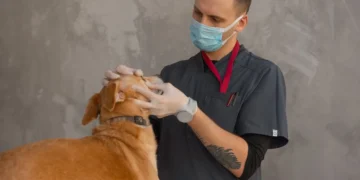
The Cranial Drawer Test – The Definitive Vet Test
The vet’s gold-standard physical exam test:
- Vet stabilises the femur with one hand
- Tries to slide the tibia forward (cranially) with the other hand
- Forward movement = positive drawer sign = CCL rupture
Sedation is often needed in tense dogs to overcome muscle spasm that masks a positive drawer in conscious patients. A negative drawer in a conscious dog does not rule out CCL disease — especially partial tears or complete tears with significant muscle guarding.
The tibial compression test is an alternative — the vet flexes the hock while feeling for cranial tibial movement. Same principle, sometimes easier to elicit.
Major Risk Factors
Breed
The most over-represented breeds:
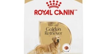
- Labrador Retriever — the standout; lifetime incidence 5-25% depending on study
- Rottweiler
- Newfoundland
- Saint Bernard
- Mastiff
- Staffordshire Bull Terrier / Pit Bull / American Staffordshire
- Boxer
- German Shepherd Dog
- Golden Retriever
- Chow Chow
- Bichon Frise (smaller-breed exception)
- Cocker Spaniel
Body Condition
Obesity is one of the strongest modifiable risk factors — mechanical loading on a degenerating ligament accelerates failure. Lean body condition (BCS 5) substantially reduces lifetime CCL risk and improves outcomes when disease occurs.
Age
Peak incidence:
- Large/giant breeds: 2-7 years (can occur in young dogs)
- Medium breeds: 5-10 years
- Small breeds: 8+ years (less common but possible)
Neuter Status
Some studies (Slauterbeck 2004) suggest early neutering in certain breeds (especially Golden Retrievers and Labradors) elevates CCL disease risk — possibly via altered skeletal development. The PuppaDogs Spay/Neuter Timing Calculator outlines the Hart et al. recommendations.
Conformation
Excessive tibial plateau angle (>30°) increases cranial tibial thrust forces and predisposes to CCL disease. This is partly heritable; selecting against high tibial plateau angles in breeding stock would reduce population incidence.
Diagnostic Workup
Physical Examination
- Lameness assessment (gait)
- Sit test
- Stifle palpation for effusion
- Cranial drawer test (under sedation if needed)
- Tibial compression test
- Range of motion
- Muscle atrophy measurement (tape around thigh)
Radiographs
Bilateral stifle radiographs typically show:
- Joint effusion — compression of the infrapatellar fat pad (“fat pad sign”)
- Osteoarthritis changes — periarticular osteophytes, soft tissue swelling
- Meniscal calcification sometimes visible (chronic disease)
- Cranial tibial drawer sign can sometimes be seen radiographically if positioned correctly
Radiographs do not directly show the CCL — they show secondary changes. The cruciate is assessed clinically, by MRI, by arthroscopy, or at surgery.
Advanced Imaging
- MRI — directly visualises the CCL and meniscus; gold standard but expensive
- Arthroscopy — visualises and treats meniscal injury in one procedure
- Stifle CT — useful for surgical planning in complex cases
Treatment Options
Surgical Treatment (Preferred For Large Athletic Dogs)
TPLO (Tibial Plateau Levelling Osteotomy)
- Current gold standard for most medium-to-giant breeds
- Cuts and rotates the tibial plateau to neutralise cranial tibial thrust
- Excellent return-to-function rates — 90%+ achieve good function
- Best long-term outcome data
- Cost: GBP 4,000-7,000 / USD 4,000-7,000 at specialist centres
TTA (Tibial Tuberosity Advancement)
- Advances the tibial tuberosity to redirect the patellar tendon force
- Widely used; similar outcomes to TPLO in many studies
- Slightly different complication profile
- Cost: similar to TPLO
Lateral Suture (Extracapsular Stabilisation)
- Heavy suture material outside the joint to stabilise
- Cheaper and simpler than TPLO/TTA
- Suitable for small-to-medium dogs (<15-20 kg)
- Inferior long-term outcome in large dogs
- Cost: GBP 1,500-3,000 / USD 1,500-3,000
Newer Procedures
- CBLO (CORA-Based Levelling Osteotomy) — variant of TPLO
- Tight Rope / SwiveLock — improved extracapsular suture material
- PAUL (Proximal Abducting Ulnar Osteotomy) — for selected cases
Conservative Management
Appropriate for:
- Small dogs <10 kg — 60-85% return to acceptable function with strict rest + NSAID + physio + weight management
- Older dogs with comorbidities precluding anaesthesia
- Bilateral disease where one stable side is essential
- Trial of conservative management before surgery in selected cases
Conservative protocol:
- Strict rest 6-8 weeks — crate rest, lead-walked toilet trips only
- NSAID — carprofen, meloxicam, Galliprant, firocoxib
- Physiotherapy / hydrotherapy — substantially improves outcomes
- Weight management to BCS 5
- Gradual return to activity over 2-3 months
Outcomes are inferior to surgery in large dogs — for athletic / large / young patients, surgery is typically recommended.
Meniscal Injury
The medial meniscus is injured in 30-80% of CCL ruptures. Treatment at surgery:
- Inspect at surgery
- Partial meniscectomy for tears
- Total meniscectomy rarely needed
- “Meniscal release” (deliberate caudal pole release) was historically performed prophylactically but is now controversial; selective meniscectomy is preferred
Untreated meniscal tears cause ongoing pain and accelerate osteoarthritis.
Bilateral Disease Reality
About 30-50% of dogs develop second-side CCL disease within 1-2 years of the first. The pattern:
- First side ruptures, surgery performed
- Recovery (8-12 weeks strict, 4-6 months full)
- Increased load on the contralateral side during recovery accelerates degeneration of an already-vulnerable second CCL
- Second side ruptures often during the rehabilitation period
Staged bilateral surgery is the standard approach. Simultaneous bilateral is occasionally chosen for owner convenience but carries substantially higher complication and recovery-burden risks — typically not recommended.
Post-Operative Care
The critical window:
- Strict rest 8-12 weeks — crate rest, lead-walked toilet only
- No jumping, stairs, running, off-leash activity
- Physiotherapy / hydrotherapy starting 2-3 weeks post-op (specialist centres)
- NSAID for pain
- Gradual return to activity weeks 12-26
- Most dogs return to good function by 4-6 months
- Full athletic function takes 6-12 months
Prevention And Risk Reduction
The best evidence-based interventions:
- Lean body condition (BCS 4-5) for life — single biggest modifiable factor
- Avoid sudden bursts of intense activity in unconditioned dogs
- Consider neuter timing for predisposed breeds (Hart et al. recommendations)
- Recognise contralateral CCL disease early if first side affected — proactive weight management, controlled exercise, watch for signs
Differentials For Hind-Limb Lameness
CCL is common but not the only diagnosis. Consider:
| Condition | Distinguishing features |
|---|---|
| Hip dysplasia | Young large breeds, often bilateral, “bunny hop” gait, positive Ortolani |
| Patellar luxation | Small breeds, “skipping” gait, palpable lateral/medial luxation |
| Hip osteoarthritis | Older dogs, hip pain on extension, X-ray confirms |
| IVDD | Neurological signs, paresis, often bilateral, back pain |
| Osteosarcoma | Older large breeds, severe pain, swelling near joint, X-ray shows bone destruction |
| Immune-mediated polyarthritis | Multiple joints, fever, lethargy, joint taps confirm |
| Soft tissue injury | Acute history, no drawer sign, often resolves |
Vet orthopaedic exam + radiographs distinguishes these.
Honest Caveats
- Pre-test probability is not diagnosis — orthopaedic exam (often under sedation) + radiographs are the diagnostic steps.
- The drawer test is not 100% reliable in conscious dogs — sedation often needed to overcome muscle guarding.
- Owner assessment of lameness severity varies; vet examination grades it consistently.
- Surgical decision-making depends on size, age, lifestyle, finances, comorbidities — specialist orthopaedic input is valuable.
- Conservative management outcomes in small dogs are reasonable but inferior to surgery in large dogs.
Conclusion
Canine CCL disease is the most common orthopaedic problem in adult dogs — predominantly degenerative, strongly breed-predisposed (Labradors especially), worsened by obesity, and presenting as hind-limb lameness with characteristic sit-test posture. Surgical stabilisation (TPLO most common for medium-to-giant breeds) typically restores best long-term function; conservative management is reasonable for small dogs and older patients. Bilateral disease is common — proactive contralateral monitoring matters. Lean body condition for life is the single best modifiable preventive intervention.
Frequently Asked Questions
How do I know if my dog has a cruciate ligament injury?
The classic signs: hind-limb lameness (variable severity), often a sudden worsening after a minor twist or jump, sitting with the affected leg extended to the side (positive ‘sit test’ – the single most useful owner sign), reduced activity, muscle atrophy of the thigh, joint swelling around the knee, sometimes clicking on movement (suggests meniscal injury). The vet’s definitive test is the CRANIAL DRAWER TEST – often under sedation to overcome muscle spasm in conscious tense dogs.
What is TPLO surgery for dogs?
TPLO (Tibial Plateau Levelling Osteotomy) is the current gold standard surgical treatment for canine CCL disease in most medium-to-giant breeds. The procedure cuts and rotates the tibial plateau to neutralise cranial tibial thrust – the force that destabilises the knee after CCL rupture. Excellent return-to-function rates (90%+) and best long-term outcome data. Typical cost GBP 4,000-7,000 / USD 4,000-7,000 at specialist centres. Recovery: 8-12 weeks strict rest, full athletic function in 6-12 months.
Can my dog recover from cruciate injury without surgery?
Conservative management is appropriate for: small dogs <10 kg (60-85% return to acceptable function with strict rest 6-8 weeks + NSAID + physiotherapy + weight management); older dogs with comorbidities precluding anaesthesia; bilateral disease where one stable side is essential. For large athletic dogs, surgery (TPLO, TTA) typically gives substantially better long-term function and is recommended where finances and patient suitability allow.
Which dog breeds are most prone to cruciate ligament rupture?
Most over-represented: Labrador Retriever (the standout, lifetime incidence 5-25%), Rottweiler, Newfoundland, Saint Bernard, Mastiff, Staffordshire Bull Terrier / Pit Bull / American Staffordshire, Boxer, German Shepherd, Golden Retriever, Chow Chow, Bichon Frise (smaller-breed exception), Cocker Spaniel. Risk compounds with obesity, athletic activity, and early neutering in some breeds. Lean body condition (BCS 5) for life is the single most important preventive intervention.
Will my other knee also rupture?
About 30-50% of dogs develop second-side cruciate disease within 1-2 years of the first. The pattern: first side ruptures, surgery, recovery; increased load on contralateral side during recovery accelerates degeneration of an already-vulnerable second CCL; second side ruptures often during rehabilitation. Staged bilateral surgery is the standard approach. Lean body condition, controlled exercise during recovery, and early recognition of second-side signs help.
How long is the recovery after cruciate surgery?
Strict rest 8-12 weeks (crate rest, lead-walked toilet trips only, no jumping/stairs/running/off-leash activity). Physiotherapy and hydrotherapy starting 2-3 weeks post-op substantially improve outcomes. Gradual return to activity weeks 12-26. Most dogs return to GOOD FUNCTION by 4-6 months. FULL ATHLETIC function takes 6-12 months. NSAID for pain management throughout the recovery period.
Related PuppaDogs Calculators
Continue building your dog’s personalised care plan with these related PuppaDogs calculators:
- Dog Pregnancy / Whelping Due-Date Calculator
- Puppy Weight Predictor (Adult Weight Calculator)
- Heatstroke Risk Calculator for Dogs
- Bloat (GDV) Risk Calculator for Dogs
- Dog Life Expectancy Calculator (Breed, Body Condition, Lifestyle)
- Spay/Neuter Timing Calculator for Dogs (Breed-Specific)
References & Further Reading
The dosing ranges and safety information on this page are drawn from the following veterinary references. Always defer to your own veterinarian and the manufacturer’s label for your specific product.
- Vasseur PB. Stifle joint. In Slatter D (ed). Textbook of Small Animal Surgery, 3rd ed. Saunders, 2003.
- Slauterbeck JR, Pankratz K, Xu KT, et al. Canine ovariohysterectomy and orchiectomy increases the prevalence of ACL injury. Clinical Orthopaedics, 2004.
- Aragon CL, Budsberg SC. Applications of evidence-based medicine: cranial cruciate ligament injury repair in the dog. Veterinary Surgery, 2005.
- Bergh MS, Sullivan C, Ferrell CL, Troy J, Budsberg SC. Systematic review of surgical treatments for cranial cruciate ligament disease in dogs. JAAHA, 2014.
- Wilke VL, Robinson DA, Evans RB, Rothschild MF, Conzemius MG. Estimate of the annual economic impact of treatment of cranial cruciate ligament injury in dogs in the United States. JAVMA, 2005.
- ACVS (American College of Veterinary Surgeons) guidelines on canine CCL disease.
- PuppaDogs. Ideal Weight & Weight Loss Calculator and Spay/Neuter Timing Calculator. puppadogs.com.
⚕️ Medical disclaimer
The information on this page is intended for educational purposes only and does not replace a hands-on veterinary examination. Drug doses depend on your dog’s complete clinical picture, concurrent medications, and the exact product formulation. Always confirm dosing with your veterinarian before administering any medication, and contact a 24-hour veterinary emergency service or animal poison control immediately if you suspect a medication overdose or adverse reaction. PuppaDogs editorial standards: every drug dose published here is cross-checked against multiple authoritative veterinary references and reviewed by the PuppaDogs Veterinary Editorial Team before publication.
