

















Quick take: Mast cell tumors (mastocytomas) are common skin cancers in dogs that can range from low‑grade, easily removable nodules to aggressive, invasive disease. Early detection, proper staging, and a tailored treatment plan—often surgery plus additional therapy—give many dogs a good quality of life and a solid chance of long‑term control.
It’s 11 p.m., and you’re scrolling through endless articles while your 8‑year‑old golden retriever, Marley, lies on the couch, his usual wagging tail now barely moving. A small, raised bump on his flank looks a bit pinker than his coat, and you notice his gums are a little paler than usual. Your heart races; the words “cancer” flash across the screen. You wonder: “Is this a mast cell tumor? How urgent is it? What can we do?”
First, breathe. Mast cell tumors are one of the most frequently diagnosed skin cancers in dogs, but they’re also a spectrum—some are low‑grade and curable with surgery alone, while others require radiation, chemotherapy, or targeted drugs. In the next few minutes we’ll walk through what mast cell tumors are, how to spot them early, what your vet will do to confirm the diagnosis, and the treatment, cost, and home‑care options that can help Marley (or any dog) get back to his playful self.
We’ll also share practical tips on feeding, monitoring, and preventing future growths, plus real‑world cost estimates and a quick‑reference guide you can print or bookmark. If at any point you feel something isn’t right—especially if your dog seems painful, the lump grows rapidly, or his appetite drops—call your vet right away.
What is a mast cell tumor (mastocytoma) in dogs?
A mast cell tumor (MCT) is a cancer that arises from mast cells, which are immune‑system cells that store histamine and other chemicals involved in allergic reactions. When these cells become malignant, they form a lump that can appear anywhere on the skin, but also in internal organs such as the spleen, liver, or gastrointestinal tract. Mast cell tumors are graded (Patnaik system) from Grade I (low grade, slow‑growing) to Grade III (high grade, aggressive). A newer two‑tier system (Ki‑67 index) helps predict behavior more accurately.
According to the American College of Veterinary Internal Medicine (ACVIM), mast cell tumors account for roughly 15–20 % of all cutaneous tumors in dogs, making them the second‑most common skin cancer after lymphoma. They can affect dogs of any age, but the median age at diagnosis is about 8 years. While some tumors stay localized, others can metastasize—spread—to the lymph nodes, liver, bone marrow, or other organs.
What causes mast cell tumors?
The exact trigger that turns a normal mast cell into a cancer cell remains unclear, but several risk factors have been identified:
- Breed predisposition: Certain breeds, including Boxers, Labrador Retrievers, Golden Retrievers, and Boston Terriers, have a higher incidence of MCTs.
- Age: Dogs over 7 years old are more likely to develop mast cell tumors.
- Genetic mutations: Mutations in the c‑KIT gene, which encodes a receptor tyrosine kinase, are found in many high‑grade tumors and drive growth. Targeted drugs (e.g., toceranib) exploit this pathway.
- Environmental factors: Chronic skin irritation, UV exposure, and possibly certain chemical exposures have been suggested, though definitive evidence is limited.
While you can’t change a dog’s genetics, knowing breed risk and monitoring older dogs closely helps catch tumors early.
Signs and symptoms
Early mast cell tumors often look like a small, raised bump that may be pink, red, or skin‑colored. They can be smooth or ulcerated and may bleed easily when touched. As the tumor grows, you might notice:
| Severity | Typical signs |
|---|---|
| Early / mild | A solitary, firm nodule; occasional itching; no pain. |
| Moderate | Rapid growth; ulceration or crusting; occasional bleeding; slight discomfort when pressed. |
| Severe | Multiple nodules; ulcerated, draining lesions; swelling of nearby lymph nodes; lethargy, loss of appetite, pale gums indicating anemia. |
Because mast cells release histamine, a tumor can cause localized itching, swelling, or a “flushing” sensation around the lesion. Systemic signs—such as vomiting, diarrhea, or bruising—may indicate that the cancer has spread or that histamine release is affecting the whole body.
When to call your vet
Call your vet today (non‑emergency):
- If you notice a new lump that is growing, changing color, or bleeding.
- If your dog shows mild itching or discomfort around a lump.
- If you observe any loss of appetite or slight lethargy that persists more than 24 hours.
Go to an emergency clinic right now:
- Sudden, severe swelling or ulceration that is rapidly expanding.
- Bleeding that won’t stop after a few minutes of gentle pressure.
- Signs of anaphylaxis (hives, facial swelling, difficulty breathing) after a lump is touched.
- Very pale gums, rapid breathing, or collapse—possible anemia from tumor spread.
Our guidance is for information only and does not replace a hands‑on veterinary exam. If you’re ever unsure, err on the side of calling your vet.
How vets diagnose a mast cell tumor
Diagnosing an MCT follows a stepwise approach:
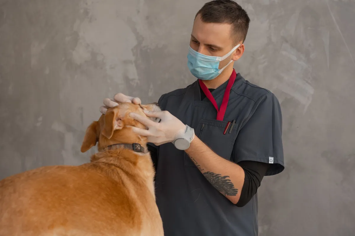
- Physical exam: Your vet will assess the lump’s size, texture, and mobility, and check nearby lymph nodes.
- Fine‑needle aspiration (FNA): A thin needle extracts cells for cytology. This often gives a quick preliminary diagnosis, but a definitive grade requires a biopsy.
- Incisional or excisional biopsy: A small piece (incisional) or the whole lump (excisional) is sent to a pathology lab. Histopathology determines the grade (Patnaik I‑III) and Ki‑67 index.
- Staging tests: Depending on the grade, the vet may recommend chest X‑rays, abdominal ultrasound, and blood work (CBC, chemistry) to look for metastasis.
- c‑KIT mutation testing: If a high‑grade tumor is suspected, testing for c‑KIT mutations helps decide whether targeted therapy (e.g., toceranib) is appropriate.
These tests give a clear picture of how aggressive the tumor is and guide the treatment plan. Your vet may also discuss the benefits of a sentinel lymph node biopsy to improve staging accuracy.
Treatment options
Medical treatment
Beyond surgery, several medical therapies are used:
- Chemotherapy: Drugs such as vincristine, cyclophosphamide, and lomustine are common for high‑grade or metastatic MCTs. They aim to shrink tumors and control spread.
- Targeted therapy: Toceranib phosphate (Palladia) and masitinib target the c‑KIT receptor, slowing tumor growth. Your vet will discuss eligibility based on mutation testing.
- Radiation therapy: External beam radiation is an option for tumors that cannot be fully removed surgically or for local recurrence.
- Supportive medications: Antihistamines (e.g., diphenhydramine) and H1/H2 blockers help control histamine release, reducing itching and swelling. Ask your vet about these options.
Supplements and supportive care
While supplements cannot cure MCTs, they can improve overall health during treatment:
- Omega‑3 fatty acids (EPA/DHA): May reduce inflammation and support immune function. Look for a high‑quality fish‑oil supplement.
- Probiotics: Helpful if chemotherapy causes gastrointestinal upset; a strain like Bifidobacterium animalis is well‑studied in dogs.
- Vitamin C: Antioxidant support is sometimes recommended, but discuss dosing with your vet.
Procedures or surgery
Complete surgical excision with clean margins (usually 2 cm of healthy tissue) is the gold standard for low‑grade MCTs. For high‑grade tumors, a wider excision plus adjunct therapy is typical. Recovery involves a 10‑14 day wound‑care period, during which you’ll keep the incision clean and limit activity.
Diet and nutrition
Nutrition plays a supportive role during and after mast cell tumor treatment. While no single diet can cure cancer, feeding a balanced, highly digestible diet helps maintain body condition, supports the immune system, and reduces stress on the liver—especially important if chemotherapy is used.
Consider the following guidelines:
| Category | Recommendations |
|---|---|
| Do feed | High‑quality protein (chicken, turkey, fish), omega‑3 enriched kibble or wet food, easily digestible carbohydrates (sweet potato, rice), and a moderate amount of healthy fats. |
| Limit | Excessive fat, especially from table scraps; high‑sodium foods that can exacerbate fluid retention. |
| Avoid | Raw meat diets unless properly balanced, as they may introduce pathogens; foods high in simple sugars, which can fuel inflammation. |
If your dog is undergoing chemotherapy, a diet low in phosphorus and high in antioxidants can help protect the liver and kidneys. Prescription therapeutic diets—such as “oncology‑support” formulas from reputable manufacturers—are formulated with added omega‑3s, antioxidants, and highly digestible proteins. Discuss options with your vet; they can tailor the feeding amount to maintain a body condition score (BCS) of 4‑5 out of 9.
Transition gradually over 5‑7 days by mixing increasing amounts of the new food with the current diet. Small, frequent meals (3‑4 times daily) are easier on a dog’s stomach during treatment. Fresh water should always be available, and avoid treats that are high in fat or sugar.
Cost and prognosis
Financial considerations vary by region, tumor grade, and treatment plan. Below are typical estimates for the United States and the United Kingdom (prices are averages; actual costs may differ based on clinic and location).
| Category | US Estimate | UK Estimate |
|---|---|---|
| Surgical excision (including anesthesia) | $1,200 – $2,500 | £800 – £1,500 |
| Biopsy and histopathology | $300 – $600 | £150 – £300 |
| Chest X‑ray & abdominal ultrasound | $200 – $400 | £100 – £250 |
| Chemotherapy (single drug course) | $1,500 – $3,000 | £1,000 – £2,200 |
| Targeted therapy (e.g., toceranib) | $2,000 – $4,500 | £1,500 – £3,500 |
| Radiation (multiple fractions) | $4,000 – $7,000 | £3,000 – £5,500 |
Prognosis depends heavily on grade and stage. Low‑grade (Patnaik I) tumors that are fully removed often have a >90 % disease‑free survival at 2 years. High‑grade (Patnaik III) tumors carry a poorer outlook, with median survival ranging from 6 months to 1 year, though targeted therapy can extend life and improve quality. Your vet will discuss individualized expectations based on pathology results.
Prevention and home care
While you can’t prevent every mast cell tumor, routine skin checks and early veterinary evaluation are key. Here are practical steps you can take:
- Perform a quick visual and tactile exam of your dog’s skin once a month—look for any new lumps, bumps, or changes in existing spots.
- Keep a photo log of any suspicious lesions; this helps track growth over time.
- Maintain a healthy weight; obesity can impair immune function.
- Use a broad‑spectrum flea and tick preventive (consult your vet for region‑appropriate products) because chronic inflammation may play a role in tumor development.
- Schedule regular wellness exams (at least annually for senior dogs) so your vet can perform fine‑needle aspirates on any concerning masses.
After surgery, follow your vet’s wound‑care instructions: keep the incision clean, prevent licking with an e‑collar, and limit strenuous activity for two weeks. Monitor for signs of infection (redness, swelling, discharge) and report them promptly.
From our vet team: “Mast cell tumors can be scary, but most owners find that early detection and a clear treatment plan make a huge difference. If you notice a new lump, have it evaluated right away. Even if the tumor is high‑grade, modern chemotherapy and targeted drugs can keep your dog comfortable and extend his happy years. We’re here to guide you through each step, from surgery to nutrition.”
Key takeaways
- Check your dog’s skin monthly for new or changing lumps, and contact your vet promptly if anything looks abnormal.
- Diagnosis requires a biopsy and grading; this determines whether surgery alone or additional therapy is needed.
- Low‑grade mast cell tumors often cure with clean surgical removal, while high‑grade tumors may need chemotherapy, radiation, or targeted drugs.
- Supportive nutrition—high‑quality protein, omega‑3s, and balanced calories—helps your dog tolerate treatment and heal faster.
- Costs can vary widely; discuss a detailed estimate and possible payment options with your veterinary clinic early on.
Myth vs. fact
Myth: All mast cell tumors are immediately life‑threatening.
Fact: Many MCTs are low‑grade and curable with surgery alone; prognosis depends on grade, stage, and location.
Myth: If a lump looks harmless, it can be ignored.
Fact: Even a small, seemingly benign nodule can be a mast cell tumor; early biopsy is the only way to know.
Myth: Home remedies can replace veterinary treatment for mast cell tumors.
Fact: While diet and supplements support overall health, definitive treatment requires veterinary intervention—surgery, chemotherapy, radiation, or targeted therapy.
Frequently asked questions
What causes mast cell tumors in dogs?
Mast cell tumors arise from genetic mutations—especially in the c‑KIT gene—combined with age‑related cellular changes. Certain breeds (Boxer, Labrador, Golden Retriever) have a higher incidence, but the exact environmental triggers remain uncertain.
How can I tell if my dog has a mast cell tumor?
The first clue is a new, firm lump that may be pink, red, or skin‑colored and can bleed when touched. A veterinarian will confirm the diagnosis with a fine‑needle aspirate and a biopsy to determine the tumor’s grade.
Are mast cell tumors cancerous?
Yes, mast cell tumors are a type of cancer. Their behavior ranges from slow‑growing, low‑grade tumors that are often curable with surgery, to aggressive, high‑grade tumors that can metastasize and require additional therapies.
What treatment options are available for mast cell tumors?
Standard treatment starts with surgical removal. Depending on the grade and stage, your vet may recommend chemotherapy (vincristine, cyclophosphamide), radiation therapy, or targeted drugs like toceranib that inhibit the c‑KIT pathway. Supportive care includes antihistamines and nutritional supplements.
How much does mast cell tumor treatment cost?
In the United States, total costs can range from $2,500 for a simple excision to over $10,000 for surgery plus chemotherapy or radiation. In the United Kingdom, similar treatment plans typically cost between £1,500 and £9,000. Your clinic can provide a detailed estimate based on your dog’s specific needs.
What is the prognosis for dogs with mast cell tumors?
Prognosis is best for low‑grade (Patnaik I) tumors that are fully removed, with over 90 % disease‑free survival at two years. High‑grade (Patnaik III) tumors have a more guarded outlook, but targeted therapies can extend survival and maintain quality of life.
Ask the PuppaDogs community
Have a question this article didn’t fully answer? Want to compare notes with other dog owners who’ve been through this? Our community forum is moderated by experienced owners and vets — and answers tend to come fast. Ask in the PuppaDogs community →
References
- American College of Veterinary Internal Medicine (ACVIM) – “Mast Cell Tumors in Dogs” guideline, 2023.
- Merck Veterinary Manual – “Mast Cell Tumors” entry, updated 2022.
- American Animal Hospital Association (AAHA) – “Canine Oncology Standards,” 2021.
- World Small Animal Veterinary Association (WSAVA) – “Guidelines for Diagnosis and Treatment of Canine Mast Cell Tumors,” 2022.
- Veterinary Cancer Society – “c‑KIT Mutations and Targeted Therapy,” 2023.
- American Veterinary Medical Association (AVMA) – “Understanding Canine Cancer,” 2023.
- University of California, Davis Veterinary Medicine – “Canine Mast Cell Tumor Staging and Prognosis,” 2022.
- Royal College of Veterinary Surgeons (RCVS) – “Surgical Management of Mast Cell Tumors,” 2021.
