













Quick take: An aortic blood clot (thrombus) is a dangerous blockage in the main artery leaving the heart. It can cause sudden weakness, pain, or loss of limb function, and it often requires urgent veterinary care. Prompt diagnosis, medical or surgical treatment, and careful home care give many dogs a good chance at recovery.
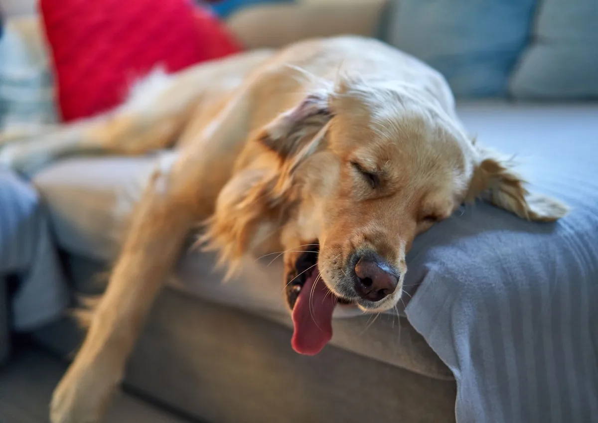
It’s 11 p.m., and your senior Golden Retriever, Max, isn’t hopping up to the door like she usually does. Instead, she lies on the rug, her hind legs trembling and her gums a pale pink. Your heart races as you Google “dog aortic clot,” hoping for a quick answer before the night deepens.
We understand that moment—those flickering worries and the urge to act. An aortic blood clot in dogs is serious, but it isn’t always a death sentence. In the next few minutes we’ll walk you through what it is, why it happens, how vets find it, treatment options, costs, and what you can do at home to give Max the best chance of a smooth recovery.
Read on for a clear, step‑by‑step guide that covers the symptoms, diagnostics, treatment pathways, diet tips, and the financial picture, all written in plain language and backed by the latest veterinary guidelines.
What is a heart (aortic) blood clot in dogs?
An aortic blood clot—also called an aortic thrombus—is a solid mass of platelets, fibrin, and red blood cells that forms inside the aorta, the large artery that carries oxygen‑rich blood from the heart to the rest of the body. In dogs, the clot typically lodges near the heart where the aorta branches into the hind‑limb arteries. When the clot blocks blood flow, the affected limbs can become weak, painful, or even necrotic.
Although any dog can develop a clot, it’s most common in senior dogs (8 years and older) and in breeds predisposed to heart disease, such as Doberman Pinschers, Boxers, and Great Danes. According to the American College of Veterinary Internal Medicine (ACVIM), aortic thrombus accounts for roughly 5–10 % of emergency cardiac cases in veterinary hospitals.
What causes it?
Clot formation is usually the result of one or more underlying problems that disturb the normal flow of blood or damage the inner lining of the aorta. The most common risk factors are:
- Heart disease: Dilated cardiomyopathy, severe mitral valve disease, or arrhythmias can cause turbulent blood flow, encouraging clot formation.
- Age: Older dogs have less elastic vessels and a higher likelihood of concurrent illnesses.
- Breed predisposition: Doberman Pinschers, Boxers, Great Danes, and German Shepherds have a higher incidence of aortic thrombus.
- Hypercoagulable states: Certain cancers, immune‑mediated diseases, or inherited clotting disorders increase clotting tendency.
- Trauma or surgery: Recent orthopedic procedures or abdominal trauma can trigger clotting cascades.
Environmental factors such as dehydration, obesity, or prolonged immobility (e.g., after a long car ride) can further tip the balance toward clot formation.
Signs and symptoms
Because the clot blocks blood flow to the hind limbs, the most noticeable signs involve the back end of the dog. Early signs may be subtle, while severe blockage can lead to life‑threatening complications.
| Severity | Typical signs |
|---|---|
| Mild | Reluctance to run, occasional stumbling, slight coldness in paws |
| Moderate | Sudden weakness or paralysis of one or both hind limbs, pale or bluish gums, pain when the affected leg is touched |
| Severe | Complete hind‑limb paralysis, severe pain, darkened or necrotic toes, rapid drop in heart rate, collapse |
Other systemic signs can include rapid breathing, a weak pulse, or a “cold” feeling in the extremities. These symptoms differ from a pulmonary embolism, which typically causes coughing, difficulty breathing, and a sudden collapse without hind‑limb weakness.
When to call your vet
Call your vet today if you notice any of the following:
- Sudden limp or weakness in one or both hind legs
- Pale, bluish, or unusually cool gums
- Visible pain when the hind limbs are touched
- Decreased appetite or lethargy that’s new or worsening
Go to an emergency veterinary hospital right now if you see any of these red‑flag signs:
- Complete paralysis of the hind limbs
- Severe, unrelenting pain (the dog cries out or cannot settle)
- Rapid heart rate combined with fainting or collapse
- Dark, mottled skin on the toes or signs of tissue death
These symptoms can indicate a life‑threatening blockage that needs immediate attention. This article is for information only and does not replace a hands‑on veterinary exam.
How vets diagnose it
Diagnosing an aortic thrombus involves a combination of history, physical exam, and imaging. The typical work‑up looks like this:
- Physical exam: The vet checks pulse quality, temperature of the hind limbs, and looks for pain or weakness.
- Blood work: A complete blood count (CBC) and chemistry panel help rule out anemia, infection, or organ dysfunction that could mimic clot symptoms.
- Imaging:
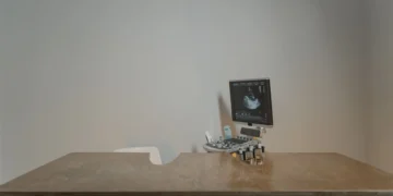
- Ultrasound (Doppler): Non‑invasive and can visualize the clot in the aorta in many cases.
- Computed tomography (CT) angiography: Provides a detailed picture of clot size and exact location, often used before surgery.
- Radiographs: May show secondary changes such as limb swelling, but are less specific.
- Electrocardiogram (ECG) and echocardiogram: Evaluate underlying heart disease that may have contributed to clot formation.
These tests together allow the veterinarian to confirm the presence of a clot, assess its severity, and plan the most appropriate treatment.
Treatment options
Medical treatment
When surgery isn’t immediately feasible, or as part of post‑operative care, vets may use medications to dissolve or prevent further clotting. Common drug classes include:
- Anticoagulants: Aspirin, clopidogrel (Plavix), or newer agents like DOACs (direct oral anticoagulants) can reduce clot propagation. Ask your vet about this option.
- Thrombolytics: Tissue plasminogen activator (tPA) or streptokinase may be given intravenously in an emergency setting to break down the clot quickly. Use only under veterinary supervision.
- Pain and anti‑inflammatory drugs: NSAIDs such as carprofen or meloxicam help control pain and inflammation, but must be used cautiously if kidney function is compromised.
Supplements and supportive care
While supplements can’t replace prescription medication, certain products may aid recovery:
- Omega‑3 fatty acids (EPA/DHA): Reduce inflammation and support vascular health. A quality fish‑oil supplement can be added after the vet confirms it’s safe.
- Vitamin C and E: Antioxidants that may help protect blood vessel walls during clot resolution.
- Probiotics: Support gut health, especially if the dog is on antibiotics.
Always discuss supplement choices with your veterinarian to avoid interactions.
Procedures or surgery
For many dogs, especially those with large or obstructive clots, surgical removal (thrombectomy) offers the best chance of restoring blood flow. The procedure typically involves:
- General anesthesia and careful monitoring of heart and blood pressure.
- Access to the aorta via a small abdominal incision.
- Removal of the clot using specialized forceps or a balloon catheter.
- Post‑operative intensive care, including pain management, anticoagulation, and fluid therapy.
Recovery in the hospital usually lasts 2–4 days, after which most dogs are discharged with strict home‑care instructions. The cost for surgery can range widely (see the Cost section below).
Diet and nutrition
Nutrition plays a pivotal role in both recovery from an aortic clot and in preventing future clot formation. While there is no “one‑size‑fits‑all” prescription diet for aortic thrombus, the following guidelines help support vascular health, reduce inflammation, and keep the heart strong.
What to feed
- Highly digestible, moderate‑protein diets: Foods with 18–22 % protein from quality sources (chicken, turkey, or fish) reduce the workload on the kidneys, which can be stressed by clot‑related inflammation.
- Omega‑3 enriched kibble or added fish oil: EPA/DHA at 0.2–0.4 % of the diet improves blood flow and lowers inflammatory markers (AAHA recommends this for dogs with cardiovascular disease).
- Limited sodium: Slightly reduced salt helps control blood pressure and fluid balance, especially in dogs with underlying heart disease.
- Antioxidant‑rich ingredients: Blueberries, carrots, and sweet potatoes provide vitamins C and E, which support vascular health.
What to limit or avoid
| Do feed | Limit | Avoid |
|---|---|---|
| High‑quality protein (chicken, fish, turkey) | Excessive treats (especially high‑fat) | Raw meat with unknown bacterial load (unless vetted) |
| Omega‑3 sources (fish oil, flaxseed) | Very high‑salt foods (processed snacks) | Grain‑heavy, low‑nutrient fillers |
| Fresh vegetables (pumpkin, green beans) | Excessive dairy (may cause GI upset) | Grapes, onions, or chocolate (toxic) |
How to transition
Switch to the new diet gradually over 5–7 days to avoid gastrointestinal upset. Start with 75 % of the current food mixed with 25 % of the new diet, then increase the new diet portion every two days. Offer small, frequent meals (3–4 times daily) for the first week after surgery to ease the digestive load.
Feeding schedule after surgery
- First 24 hours: Offer a bland diet such as boiled chicken and rice in small amounts every 4–6 hours.
- Days 2‑5: Transition to the prescribed therapeutic diet (if any) mixed with the bland diet.
- After day 5: Move to the maintenance diet outlined above, keeping meal portions consistent to maintain a healthy weight.
Hydration is equally important. Fresh water should be available at all times, and low‑sodium broths can be added to meals if the dog is reluctant to drink.
For owners seeking a ready‑made solution, many veterinary nutritionists recommend a “cardiac‑support” diet (e.g., Hill’s Prescription Diet k/d or Royal Canin Cardiac). These formulas are formulated with reduced sodium and enhanced taurine, but the final choice should be guided by your vet’s assessment of your dog’s overall health.
Cost and prognosis
Financial considerations are a real part of any serious medical episode. Below is an estimate of typical expenses in the United States and the United Kingdom. Prices can vary by region, clinic, and the complexity of the case.
| Service | US (USD) | UK (GBP) |
|---|---|---|
| Initial exam & blood work | $150–$300 | £80–£150 |
| Ultrasound (Doppler) or CT angiography | $300–$800 (US), $250–$600 (CT) | £200–£500 |
| Surgical thrombectomy (incl. anesthesia) | $3,000–$6,500 | £2,000–£4,500 |
| Post‑op ICU care (48 hrs) | $500–$1,200 | £300–£700 |
| Medications (anticoagulants, pain meds) | $100–$400 | £70–£200 |
| Follow‑up visits & labs (first 2 months) | $200–$400 | £120–£250 |
Overall, owners can expect to spend anywhere from $4,000 to $10,000 (or £3,000–£7,500) for a full diagnostic and surgical episode, plus ongoing medication and monitoring costs.
Prognosis depends on clot size, location, and the dog’s overall health. Dogs that receive prompt treatment—especially surgical removal—have a reported survival rate of 60–80 % and can return to a near‑normal life within 2–3 months. Those managed medically alone have a higher risk of recurrence, but many still live comfortable lives with long‑term anticoagulation.
Prevention and home care
Preventing a repeat clot starts with managing the underlying risk factors:
- Regular heart screening: Annual echocardiograms for senior dogs or breeds prone to cardiomyopathy can catch problems early. Use our heart health calculators to track heart rate trends.
- Maintain a healthy weight: Obesity strains the heart and promotes inflammation. Aim for a body condition score (BCS) of 4–5 on a 9‑point scale.
- Exercise wisely: Short, frequent walks keep circulation moving without over‑exerting a recovering heart.
- Hydration: Encourage water intake, especially on hot days, to prevent blood thickening.
- Monitor for early signs: Keep an eye on limb strength, gum color, and appetite. A quick check of the hind limbs each morning can catch subtle changes.
- Medication compliance: If your vet prescribes an anticoagulant, give it exactly as directed and never skip doses.
After surgery, the home‑care routine includes:
- Restricting activity for 2–3 weeks (no running, jumping, or stairs).
- Warming the affected limbs with a gentle heat pack (not hot) for 10 minutes twice daily to promote circulation.
- Checking the incision site for swelling, redness, or discharge.
- Providing a soft, non‑slippery surface for the dog to lie on.
Follow‑up appointments every 2–4 weeks during the first three months allow the vet to adjust medications and ensure the clot does not recur.
From our vet team: “Aortic thrombus is frightening, but early detection and a collaborative treatment plan give many dogs a second chance. Keep a close eye on your senior dog’s gait and gum color, and don’t hesitate to call us if anything feels off.”
Key takeaways
- Aortic blood clots block blood flow to the hind limbs and require urgent veterinary attention.
- Common risk factors include heart disease, older age, and breeds like Doberman Pinscher and Boxers.
- Symptoms range from mild limp to complete paralysis; red‑flag signs demand immediate emergency care.
- Diagnosis relies on physical exam, blood work, and imaging such as ultrasound or CT angiography.
- Treatment options include anticoagulants, thrombolytics, and surgical thrombectomy; early intervention improves survival.
- Post‑op recovery involves a low‑stress diet rich in omega‑3s, limited sodium, and careful activity restriction.
- Regular heart screening, weight management, and consistent medication adherence are key to preventing recurrence.
Myth vs. fact
Myth: Aortic clots only happen in very large dogs.
Fact: While larger breeds have a higher incidence, any senior dog with heart disease can develop an aortic thrombus.
Myth: Surgery is always the only cure.
Fact: Medical management with anticoagulants can be effective for small clots or dogs not suited for surgery, though recurrence risk may be higher.
Myth: Once a clot is gone, the dog is out of danger.
Fact: Ongoing monitoring and preventive care are essential because clotting disorders can recur.
Frequently asked questions
What does an aortic blood clot feel like for a dog?
Dogs can’t tell us directly, but they often show sudden weakness, pain when the hind limbs are touched, or a reluctance to stand. You may also notice pale or bluish gums and a cool feeling in the paws.
How quickly can a heart clot cause death in a dog?
In severe cases, a large clot can cut off blood flow within minutes to hours, leading to rapid tissue death and shock. Prompt veterinary care dramatically reduces the risk of fatality.
Are there any non‑surgical treatments for aortic clots in dogs?
Yes. Anticoagulant drugs (like clopidogrel), thrombolytic agents (tPA), and aggressive pain management can be used, especially when the clot is small or the dog isn’t a surgical candidate.
What diagnostic tests confirm an aortic clot in a dog?
Veterinarians typically start with an ultrasound (Doppler) to visualize the clot, followed by CT angiography for detailed mapping. Blood panels and echocardiograms help identify underlying heart disease.
Can a dog live a normal life after aortic clot removal?
Many dogs return to their regular activities within 2–3 months after successful surgery and proper rehabilitation. Ongoing heart monitoring and lifestyle adjustments help maintain long‑term health.
What medications are used to dissolve aortic clots in dogs?
Thrombolytics such as tissue plasminogen activator (tPA) are used in emergency settings, often alongside anticoagulants like clopidogrel or aspirin to prevent new clot formation.
Ask the PuppaDogs community
Have a question this article didn’t fully answer? Want to compare notes with other dog owners who’ve been through this? Our community forum is moderated by experienced owners and vets — and answers tend to come fast. Ask in the PuppaDogs community →
References
- American College of Veterinary Internal Medicine (ACVIM) 2023 Consensus Statement on Canine Cardiovascular Disease.
- AAHA 2022 Guidelines for the Management of Canine Heart Disease.
- Merck Veterinary Manual, “Aortic Thrombus” entry.
- Veterinary Clinics of North America: Small Animal Practice, “Thromboembolic Disease in Dogs,” 2021.
- World Small Animal Veterinary Association (WSAVA) Nutritional Recommendations for Dogs with Cardiovascular Disease.
- University of California, Davis Veterinary Medicine, “Thrombosis and Anticoagulant Therapy in Dogs,” 2022.
- Royal College of Veterinary Surgeons (RCVS) Guidelines on Surgical Thrombectomy in Companion Animals, 2020.
