












Quick take: Cherry eye, or eyelid protrusion, is a painless swelling of the third eyelid’s gland that slides out of its normal spot. It’s most common in young, small‑to‑medium dogs, especially certain breeds. Surgery is the standard cure, but early detection, proper eye care, and a balanced diet can help prevent recurrence.
It’s 9 p.m., you’re scrolling through pet forums, and your 2‑year‑old Boston Terrier, Milo, is blinking oddly and has a bright red bump hanging from the inner corner of his left eye. The swelling looks like a tiny cherry, and your heart jumps—“Is this an emergency?” you wonder. You’re not alone; many owners first notice cherry eye because their dog suddenly looks “red‑eyed” or rubs the area with a paw. While the sight can be unsettling, the condition is usually not painful, and with the right steps you can get Milo back to his playful self quickly.
We get it: you want to know what cherry eye really is, why it happens, how serious it can get, and what you’ll need to do—now and after treatment. Below we break down the anatomy, risk factors, signs, diagnosis, treatment options, diet tips, costs, and prevention strategies. By the end you’ll have a clear plan and know exactly when a vet visit is urgent.
What is eyelid protrusion (cherry eye) in dogs?
Cherry eye is the popular name for a prolapsed third‑eyelid gland (also called the nictitating membrane gland). The third eyelid is a thin, protective flap that slides across the eye’s surface, and it contains a small tear‑producing gland. When the connective tissue that holds the gland in place weakens, the gland can slip out of its socket and appear as a smooth, pink‑red, round mass—much like a cherry.
While the exact prevalence varies, the American College of Veterinary Ophthalmology (ACVO) estimates that cherry eye affects roughly 2–3 % of dogs, with a higher incidence in certain breeds. It most often shows up in puppies or young adults, but older dogs can develop it too, especially if they have a history of eye problems.
What causes cherry eye in dogs?
The underlying cause is a combination of genetics, anatomy, and occasionally trauma. Below are the main risk factors:
| Factor | Explanation |
|---|---|
| Genetic predisposition | Weak connective tissue in the gland’s supporting ligaments is inherited in many breeds. |
| Breed anatomy | Some breeds have a naturally larger or more mobile third‑eyelid gland. |
| Age | Puppies and young dogs have more elastic tissue, making prolapse easier. |
| Eye irritation or trauma | Frequent rubbing, scratches, or infections can aggravate the gland. |
| Hormonal influences | Hormonal changes (e.g., during spaying) may affect ligament strength, though evidence is limited. |
Breeds most commonly reported with cherry eye include:
- Bulldogs (English and French)
- Boston Terriers
- Cavalier King Charles Spaniels
- Shih Tzus
- American Cocker Spaniels
- Beagles
- Miniature Schnauzers
Signs and symptoms
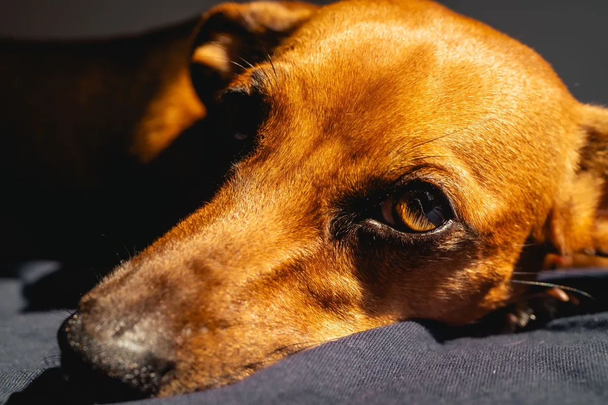
Owners usually notice a sudden, fleshy, pink‑red bump in the inner corner of the eye. The swelling can be unilateral or, less often, affect both eyes. Below is a quick guide to what you might see, from mild to severe.
| Severity | Typical signs |
|---|---|
| Mild | Small, smooth, cherry‑colored mass; mild tearing; occasional blinking. |
| Moderate | Larger mass; increased tear production; eye may look “wet” or glossy; occasional pawing. |
| Severe | Very large protrusion; chronic irritation; conjunctivitis (redness of the white of the eye); possible corneal ulcer if untreated. |
Key things to watch for:
- Persistent swelling that doesn’t shrink on its own.
- Excessive tearing or mucoid discharge.
- Rubbing the eye with a paw or leaning against furniture.
- Signs of discomfort such as squinting or reluctance to play.
When to call your vet
Call your regular vet today if you notice:
- A new or enlarging red bump on the inner eye corner.
- Excessive tearing, discharge, or squinting.
- Any sign of pain (whining, pawing at the eye).
Go to an emergency veterinary hospital right now if you see:
- Sudden loss of vision or a cloudy cornea.
- Bleeding from the eye.
- Severe swelling that blocks the eye’s surface.
This information is for educational purposes only and is not a substitute for professional veterinary care.
How vets diagnose cherry eye
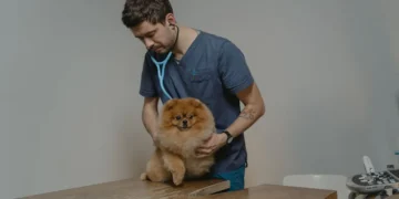
Diagnosis is usually straightforward, but a thorough exam rules out other eye problems that can look similar, such as entropion (inward‑rolling eyelid) or conjunctivitis.
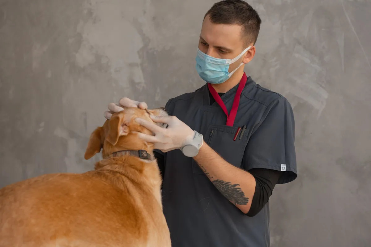
- History & visual inspection: The vet will ask when the swelling appeared, any recent eye irritation, and will gently examine the eye with a slit‑lamp or ophthalmoscope.
- Fluorescein stain: A drop of dye helps highlight corneal scratches or ulcers that may develop if the eye is rubbed excessively.
- Palpation: The vet may gently press the gland back into place to see if it’s still mobile, which confirms the diagnosis.
- Schirmer tear test (optional): Measures tear production; low tear output can indicate a compromised gland, guiding treatment decisions.
Because cherry eye can occasionally be confused with other eyelid disorders, a skilled ophthalmologist may use a specialized eye chart or even an ultrasound in rare cases.
Treatment options
Medical treatment
Medical management can temporarily reduce swelling but rarely cures the underlying prolapse. Options include:
- Topical anti‑inflammatories: Eye drops such as prednisolone acetate can lessen irritation.
- Lubricating artificial tears: Keep the eye moist while the gland is out of place.
- Systemic antibiotics: If a secondary bacterial infection is suspected.
These measures are supportive; they do not prevent the gland from slipping back out. Your vet will likely advise you to discuss surgical correction as the definitive solution.
Supplements and supportive care
While no supplement can replace surgery, some adjuncts may help overall eye health:
- Omega‑3 fatty acids (EPA/DHA): Anti‑inflammatory properties that support tear film quality. Look for a high‑quality fish‑oil supplement approved for dogs.
- Vitamin A‑rich foods: Carrots, sweet potatoes, and liver support ocular surface health.
- Probiotics: Maintaining a healthy gut can indirectly benefit immune function, which matters if your dog has recurrent eye infections.
Always ask your vet before adding any supplement, especially if your dog is on medication.
Procedures or surgery
Surgery is the gold‑standard treatment and has a success rate of 85‑95 % when performed by a board‑certified ophthalmologist. The most common techniques are:
- Pre‑auricular gland repositioning (Peter’s technique): The gland is sutured to the peri‑ocular tissue without removing any tissue.
- Lock‑and‑key (Hood’s technique): The gland is tucked into a pocket and secured with a small suture “key.”
- Excision (rare): The gland is removed only if it’s severely damaged; this can lead to dry eye, so it’s a last resort.
All procedures are done under general anesthesia, typically as a short, same‑day surgery. Recovery is usually smooth, but follow‑up care is essential to avoid recurrence.
Diet and nutrition
While diet alone won’t fix a prolapsed gland, proper nutrition supports overall eye health, immune function, and tissue integrity. Below are guidelines to help your dog recover faster and reduce the chance of recurrence.
| Do feed | Limit | Avoid |
|---|---|---|
| High‑quality protein (chicken, turkey, fish) | Excessive grain fillers | Highly processed “by‑product” kibble |
| Omega‑3 rich foods (salmon, sardines, fish oil) | High‑salt treats | Excessive raw bones that can splinter |
| Vitamin A sources (carrots, sweet potatoes, liver) | Excessive dairy | Artificial additives and preservatives |
Key points for a cherry‑eye‑friendly diet:
- Balanced protein: Supports the connective tissue that holds the gland in place. Aim for at least 22 % protein on a dry‑matter basis.
- Omega‑3 supplementation: 1 % of the diet as fish oil can reduce inflammation and improve tear film quality. Ask your vet for a dose based on weight.
- Hydration: Adequate water intake keeps tear production steady. Fresh water should be available at all times.
- Prescription therapeutic diets: Some brands offer “ocular health” formulas enriched with antioxidants (vitamin E, lutein) that may benefit dogs with chronic eye issues. Discuss options with your vet; we’re not endorsing any specific brand.
- Feeding schedule: Small, frequent meals can be easier on a recovering eye, especially if the dog is less inclined to eat a large bowl after surgery.
Transition any new food over 7–10 days to avoid gastrointestinal upset: mix 25 % new food with 75 % old, then gradually increase the new portion.
Cost and prognosis
Cherry eye surgery costs vary by region, clinic type, and whether any complications arise. Below are typical price ranges (estimates, before insurance):
| Region | Typical cost (USD) | Typical cost (GBP) |
|---|---|---|
| United States (general practice) | $300–$800 | £230–£620 |
| United Kingdom (private clinic) | £250–£700 | $340–$950 |
| Australia (specialist referral) | AUD 400–AUD 1,200 | $280–$840 |
Most pet insurance plans cover the surgery if it’s deemed medically necessary, though pre‑authorization is often required. Out‑of‑pocket costs can rise if post‑op medications, follow‑up exams, or complications (e.g., infection) occur.
Prognosis after successful repositioning is excellent. Studies cited by the American College of Veterinary Ophthalmologists (ACVO) show a 90 % long‑term success rate when the gland is correctly secured. Recurrence can happen, especially if the original suture loosens; a second surgery usually resolves the issue.
Prevention and home care
While genetics play a big role, you can lower the odds of cherry eye returning by maintaining eye health and monitoring your dog’s environment.
- Regular eye checks: Look for early swelling during weekly grooming. A quick glance at the inner corner can catch a prolapse before it enlarges.
- Keep the eye clean: Use a damp, lint‑free cloth to wipe away discharge. Avoid harsh chemicals; sterile saline is safest.
- Control allergies and irritants: Seasonal pollen or dust can cause itching, prompting the dog to rub the eye.
- Protect from trauma: Trim long hair around the eyes and discourage rough play that might lead to scratches.
- Nutrition: Continue the eye‑supportive diet long‑term, especially if your dog has a history of eye problems.
- Follow‑up visits: Your vet will likely schedule a re‑check 7–10 days post‑op, then again at 4–6 weeks to ensure the gland stays in place.
For owners who love tracking expenses, our cost calculator can help you estimate surgery and medication fees in your region.
Vet’s note
From our vet team: Cherry eye is rarely an emergency, but it does need prompt veterinary attention to avoid secondary infections or chronic dry eye. If you catch it early, the surgery is quick and recovery is usually uneventful. Keep a close eye (pun intended) on your dog’s eyelids, especially after spaying or during allergy season, and don’t hesitate to reach out to us if you’re unsure about any symptom.
Key takeaways
- Cherry eye is a prolapsed third‑eyelid gland that appears as a smooth, red “cherry” at the inner eye corner.
- Breeds such as Bulldogs, Boston Terriers, and Cavalier King Charles Spaniels are most at risk, often showing signs in puppyhood.
- Prompt veterinary evaluation is essential; surgery offers a 90 % success rate and is the definitive cure.
- Supportive nutrition—high‑quality protein, omega‑3s, and vitamin A—helps tissue health and may reduce recurrence.
- Post‑op care includes gentle eye cleaning, anti‑inflammatory drops, and a follow‑up exam within two weeks.
- If you notice swelling, excessive tearing, or any sign of pain, contact your vet today; go to an emergency clinic if vision is lost or the eye looks cloudy.
Myth vs. fact
Myth: Cherry eye is always painful and your dog will cry out.
Fact: Most dogs with cherry eye experience little to no pain; the main sign is a visible swelling and increased tearing.
Myth: Surgery is optional and the gland will heal on its own.
Fact: The gland does not re‑attach without intervention; without surgery, it often remains protruded, leading to chronic irritation and possible dry eye.
Myth: Cherry eye and entropion are the same condition.
Fact: Cherry eye is a gland prolapse, while entropion is an inward‑rolling eyelid that can cause the lashes to rub the cornea—different problems requiring different treatments.
Frequently asked questions
What is cherry eye in dogs?
Cherry eye is the common name for a prolapsed third‑eyelid gland that appears as a pink‑red, cherry‑shaped mass at the inner corner of the eye.
How can I tell if my dog has cherry eye?
The hallmark sign is a smooth, fleshy bump in the inner eye corner, often accompanied by increased tearing or occasional pawing at the eye.
Is cherry eye dangerous for my dog?
On its own, cherry eye isn’t life‑threatening, but it can lead to chronic irritation, infection, or dry eye if left untreated.
What are the treatment options for cherry eye?
Medical eye drops can soothe irritation, but surgery to reposition the gland (Peter’s or Hood’s technique) is the definitive cure with a high success rate.
How much does cherry eye surgery cost?
In the United States, expect $300–$800 for a standard procedure; costs vary by region and may be partially covered by pet insurance.
Can cherry eye come back after surgery?
Recurrence is possible, especially if sutures loosen. A second surgery typically resolves repeat cases, and proper post‑op care reduces the risk.
How long is the recovery after cherry eye surgery?
Most dogs recover within 7–10 days, with full activity resuming after two weeks. Follow‑up exams ensure the gland stays in place.
Can diet help prevent cherry eye?
While diet can’t change genetics, a balanced diet rich in omega‑3 fatty acids, high‑quality protein, and vitamin A supports overall eye health and tissue strength.
Ask the PuppaDogs community
Have a question this article didn’t fully answer? Want to compare notes with other dog owners who’ve been through this? Our community forum is moderated by experienced owners and vets — and answers tend to come fast. Ask in the PuppaDogs community →
References
- American College of Veterinary Ophthalmologists (ACVO). “Third Eyelid Gland Prolapse (Cherry Eye) Guidelines.” 2023.
- American Veterinary Medical Association (AVMA). “Eye Health in Dogs.” 2022.
- Merck Veterinary Manual. “Cherry Eye (Third Eyelid Gland Prolapse).” 2021.
- American Animal Hospital Association (AAHA). “Canine Ophthalmology Standards.” 2022.
- Cornell University College of Veterinary Medicine. “Eye Anatomy and Common Disorders.” 2023.
- World Small Animal Veterinary Association (WSAVA). “Nutritional Recommendations for Ocular Health.” 2022.
- Veterinary Ophthalmology (Journal). “Outcomes of Surgical Techniques for Cherry Eye.” 2020.
- PetMD. “Cherry Eye in Dogs: Causes, Diagnosis, and Treatment.” 2023.


