














Quick take: Excess blood cells in a dog’s eye—known as hyphema—are usually a sign of bleeding inside the eye. It can range from a harmless spot to a serious emergency, depending on the cause and how much vision is affected. Prompt veterinary care, proper diagnosis, and tailored treatment give most dogs a good chance of recovery.
It’s 11 p.m., and your golden‑crowned retriever, Bella, is lying on the couch with one eye looking bright red, almost like a tiny pool of blood. You’ve never seen anything like it, and a quick Google search later you’re scrolling through pictures of “red eye in dogs.” Your heart races—could this be something dangerous? You’re not alone; many owners discover a sudden red spot in their dog’s eye and wonder if it’s an emergency.
First, take a breath. A red eye can mean many things, from a simple irritation to a deeper bleed called hyphema. In this article we’ll explain what hyphema is, why it happens, what to look for, how vets figure it out, and what treatments—medical or surgical—might be recommended. We’ll also cover costs, diet tips, and ways to help prevent future eye bleeding.
By the end you’ll know when to call your vet right away, what questions to ask at the appointment, and how to support your dog’s recovery at home. Let’s start with the basics.
What is excess blood cells in the eye in dogs?
Excess blood cells in the eye, medically called hyphema, is a collection of red blood cells that pools in the front chamber of the eye (the anterior chamber) or sometimes in the back (the posterior chamber). The eye is filled with clear fluid called aqueous humor; when blood leaks into this space, it can turn the fluid pink or dark red, giving the eye a “blood‑filled” appearance.
Hyphema is not a disease itself but a symptom of an underlying problem—much like a cough signals a respiratory issue. It can affect dogs of any age, but it’s more common in senior dogs and certain breeds with predispositions to eye trauma or vascular disease.
In the United States, hyphema accounts for roughly 1–2 % of all ocular emergencies seen at veterinary teaching hospitals (AAHA 2022). While relatively uncommon, it’s a condition that warrants prompt veterinary attention because it can threaten vision or indicate a systemic disease.

What causes it?
Blood can enter the eye for many reasons. The most common causes in dogs include:
- Trauma: A bump, scratch, or blunt force—like a ball hitting the face—can rupture tiny blood vessels in the iris or ciliary body.
- Hypertension (high blood pressure): Especially in older, small‑breed dogs, chronic high pressure can cause delicate vessels to leak.
- Coagulation (clotting) disorders: Conditions like hemophilia, von Willebrand disease, or the use of anticoagulant medications can make bleeding more likely.
- Eye tumors: Melanomas or other intra‑ocular neoplasms can erode vessels, leading to bleeding.
- Inflammation (uveitis): Inflammation of the uveal tract can damage vessels and cause blood to seep into the eye.
- Systemic infections: Tick‑borne diseases (e.g., Ehrlichiosis) or other infections may affect the eye’s vasculature.
Less common triggers include retinal detachment, severe allergic reactions, or iatrogenic causes (e.g., a needle accident during a routine eye exam).
Signs and symptoms
Owners usually notice a visible red or pink discoloration, but other signs can develop as the condition progresses. Below is a quick guide to what you might see.
| Severity | Signs you may notice |
|---|---|
| Mild | Small, localized red spot in the white of the eye; slight cloudiness; mild squinting. |
| Moderate | Larger area of blood‑tinged fluid; noticeable cloudiness; frequent blinking; reluctance to play or run. |
| Severe | Eye appears dark red or black; marked pain (pawing at the eye, vocalizing); loss of vision; swelling of the eye (proptosis). |
Other accompanying clues include:
- Redness of the sclera (white of the eye) beyond the blood spot.
- Discharge—clear, watery, or slightly bloody.
- Changes in pupil size or shape.
- Behavioral shifts: decreased activity, reluctance to chase toys, or sensitivity to light.
When to call your vet
Call your vet today if you notice any of the following:
- A new red spot or any blood in the eye, even if it’s small.
- Swelling, pain (your dog pawing at the eye), or signs of vision loss.
- Accompanying symptoms such as vomiting, lethargy, or a sudden change in behavior.
Go to an emergency veterinary hospital right now if you see:
- Rapidly spreading redness that fills the whole eye.
- Severe pain (continuous whimpering, shaking head, or intense squinting).
- Signs of head trauma, uncontrolled bleeding, or a sudden loss of vision.
These are urgent signs that could threaten your dog’s sight or indicate a life‑threatening systemic issue. Remember, this article is for information only; it does not replace a hands‑on veterinary exam.
How vets diagnose it
Diagnosing hyphema starts with a thorough history and physical exam. Your vet will ask about recent injuries, medications, and any underlying health problems.
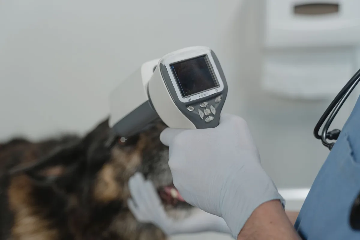
- Ophthalmic examination: Using a slit‑lamp microscope, the vet evaluates the front of the eye, checks the size and location of the blood, and looks for signs of trauma or inflammation.
- Tonometry: Measures intra‑ocular pressure to rule out glaucoma, which can coexist with hyphema.
- Ultrasound (ultrasonography): An eye ultrasound helps visualize structures behind the iris, identifying retinal detachment or intra‑ocular masses.
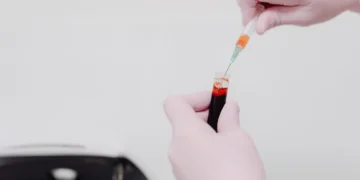
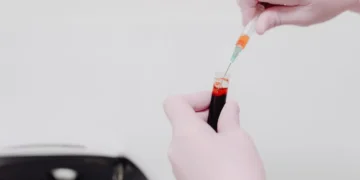
- Blood work: Complete blood count (CBC) and coagulation profile detect clotting disorders or systemic infections.
- Blood pressure measurement: Detects hypertension, especially in older small‑breed dogs.
- Advanced imaging (CT or MRI): In rare cases, when a tumor is suspected, cross‑sectional imaging may be recommended.
These tests give a clear picture of why the blood entered the eye and guide the appropriate treatment plan.
Treatment options
Medical treatment
Most cases start with medication to control inflammation, reduce intra‑ocular pressure, and prevent further bleeding. Common drug classes include:
- Topical corticosteroids: Such as prednisolone acetate, to calm inflammation.
- Systemic anti‑inflammatories: NSAIDs like carprofen may be prescribed for pain and swelling.
- Antifibrinolytic agents: Tranexamic acid helps stop bleeding by stabilizing clots (ask your vet about this option).
- Blood pressure drugs: ACE inhibitors or calcium‑channel blockers if hypertension is identified.
- Antibiotics: If an infection is suspected, broad‑spectrum antibiotics may be used.
These medications are typically given for several weeks, with frequent re‑checks to monitor the eye’s appearance and your dog’s comfort.
Supplements and supportive care
While supplements can’t replace proper medical therapy, they may aid recovery when used alongside prescribed drugs:
- Omega‑3 fatty acids (EPA/DHA): Found in fish oil, they support ocular inflammation reduction.
- Vitamin C: An antioxidant that may help strengthen blood vessel walls, but should be used only under vet guidance.
- Probiotics: If your dog is on long‑term antibiotics, a probiotic can maintain gut health.
Always discuss supplement use with your veterinarian to avoid interactions with prescribed medications.
Procedures or surgery
If the hyphema is large, does not clear, or is caused by an underlying tumor, surgery may be recommended. Options include:
- Anterior chamber washout: The vet flushes the blood out of the front of the eye, often combined with a temporary intra‑ocular lens implant.
- Laser photocoagulation: A laser seals leaking vessels, useful for small, localized bleeds.
- Enucleation (removal of the eye): Rare, reserved for eyes that are blind and painful, or when a malignant tumor cannot be salvaged.
Post‑operative care typically involves topical antibiotics, anti‑inflammatories, and protective eye patches. Recovery can take 2–4 weeks, during which activity should be limited to prevent re‑injury.
Diet and nutrition
While no single diet cures hyphema, feeding a balanced, anti‑inflammatory diet can support overall eye health and aid recovery. Here are some nutrition guidelines to consider:
- High‑quality protein: Easily digestible protein (e.g., chicken, turkey, or fish) helps maintain tissue repair without overloading the kidneys.
- Omega‑3 fatty acids: Aim for a diet that provides at least 1 % EPA/DHA of total calories. Commercial diets labeled “Joint Health” or “Cardiovascular Support” often contain adequate levels.
- Antioxidant‑rich foods: Vitamin E (found in sunflower oil) and lutein (present in green leafy vegetables) protect ocular cells from oxidative stress.
- Low sodium: Reducing sodium helps control blood pressure, especially in hypertensive dogs.
- Avoid excessive vitamin K antagonists: Some raw‑food diets can be low in vitamin K, which is essential for clotting. If you feed raw, discuss supplementation with your vet.
Prescription therapeutic diets may be advised if your dog has concurrent conditions:
| Dietary action | Recommendation |
|---|---|
| Do feed | High‑quality, moderate‑fat commercial kibble with added omega‑3; cooked lean protein; fresh vegetables (pumpkin, carrots). |
| Limit | High‑salt treats, processed foods, and excessive bone meal. |
| Avoid | Very high‑protein “raw” diets without balanced vitamin K; foods high in vitamin A (liver) which can exacerbate inflammation. |
Transition any new diet gradually over 5–7 days to avoid gastrointestinal upset. Offer smaller, frequent meals during recovery to keep energy levels steady without over‑loading the digestive system.
Cost and prognosis
Financial considerations vary widely by region, clinic, and treatment choice. Below are typical cost ranges (USD and GBP) based on 2024 data from the AAHA and veterinary practice surveys.
| Service | US estimate | UK estimate |
|---|---|---|
| Initial eye exam & diagnostics | $150–$300 | £120–£250 |
| Blood work & blood pressure check | $80–$150 | £70–£130 |
| Medical management (drugs for 4 weeks) | $100–$250 | £80–£200 |
| Anterior chamber washout surgery | $1,200–$2,500 | £900–£1,800 |
| Laser photocoagulation | $800–$1,500 | £600–£1,200 |
| Post‑op follow‑up (3 visits) | $150–$300 | £120–£250 |
Pet insurance often covers a portion of diagnostic tests and surgical fees, but coverage varies. Check your policy’s ophthalmology limits and pre‑authorization requirements. The PuppaDogs cost calculator can help you estimate out‑of‑pocket expenses based on your location.
Prognosis depends on the underlying cause, the size of the bleed, and how quickly treatment begins. For traumatic hyphema caught early, many dogs regain full vision within a month. Hypertensive or tumor‑related cases may have a guarded prognosis, and some dogs may retain permanent vision loss despite therapy. Your vet will discuss the likely outcome based on your dog’s specific situation.
Prevention and home care
While you can’t control every accident, several everyday habits can lower the risk of eye bleeding:
- Regular veterinary check‑ups: Annual exams (or more often for senior dogs) catch hypertension and clotting disorders early.
- Protect the eyes during play: Use dog‑safe toys that don’t have sharp edges, and supervise high‑energy games that could lead to head trauma.
- Maintain a healthy weight: Obesity contributes to hypertension; a balanced diet and routine exercise help keep blood pressure in check.
- Monitor blood pressure: If your dog is a breed prone to hypertension (e.g., Miniature Schnauzers, Poodles), ask your vet about periodic BP checks.
- Stay up‑to‑date on vaccinations and parasite control: Prevent infections like Ehrlichiosis that can affect the eye’s blood vessels.
- Gentle eye cleaning: If your dog has a mild irritation, a warm, damp cloth can soothe the area—never apply human eye drops without veterinary approval.
If you notice a small red spot but no pain, you can gently rinse the eye with sterile saline (available at pharmacies) to remove surface debris. However, do not attempt to “stop the bleeding” with pressure or home remedies; that can worsen the situation.
From our vet team: “When a dog presents with hyphema, our first priority is to find the root cause—whether it’s a simple bump or a hidden systemic disease. Most owners are relieved to learn that early intervention often leads to full recovery, but the key is acting quickly. If you’re ever unsure, a brief call to your clinic for advice is better than waiting for the next morning.”
Key takeaways
- Hyphema is a bleed inside the eye; it can signal anything from minor trauma to serious systemic disease.
- Call your vet today for any new red spot, pain, or vision changes; go to an emergency clinic if the eye looks dark, swollen, or the dog is in obvious distress.
- Diagnosis involves a thorough eye exam, ultrasound, blood work, and sometimes blood pressure measurement.
- Treatment may be medical (anti‑inflammatories, blood pressure meds) or surgical (washout, laser), depending on severity.
- Feeding a balanced diet rich in omega‑3s, antioxidants, and moderate protein supports healing and overall eye health.
- Regular vet visits, weight management, and safe play habits are the best ways to prevent future eye bleeding.
Myth vs. fact
Myth: All red eyes are caused by blood inside the eye.
Fact: Redness can result from irritation, infection, glaucoma, or hyphema; a vet exam determines the exact cause.
Myth: Home remedies like applying tea bags will stop eye bleeding.
Fact: Applying pressure or unapproved substances can worsen hyphema; only a vet‑approved treatment plan is safe.
Myth: Once the blood clears, the problem is solved.
Fact: Even after the visible blood disappears, underlying issues (e.g., hypertension) may still need management to prevent recurrence.
Frequently asked questions
What does blood in a dog’s eye indicate?
Blood in the eye (hyphema) indicates that a vessel inside the eye has ruptured, allowing red blood cells to mix with the clear fluid. It can be caused by trauma, high blood pressure, clotting disorders, or tumors.
Is hyphema painful for dogs?
Yes, many dogs experience discomfort. Pain may manifest as pawing at the eye, squinting, or vocalizing, especially if the bleed is large or pressure builds up inside the eye.
Can a dog recover from eye bleeding without surgery?
Most dogs with mild to moderate hyphema recover with medical management—anti‑inflammatories, blood‑pressure control, and careful monitoring. Surgery is reserved for large, persistent bleeds or when an underlying tumor is present.
How quickly should I see a vet if my dog has a red eye?
Contact your vet within the same day for any new red spot or eye discomfort. If the eye looks dark, the dog is in pain, or vision loss is suspected, seek emergency care immediately.
What are the long‑term effects of eye hemorrhage in dogs?
Potential long‑term effects include chronic inflammation, cataract formation, glaucoma, or permanent vision loss, especially if the underlying cause isn’t addressed.
Are there any home remedies for a dog’s eye bleed?
Home care is limited to gentle cleaning with sterile saline. No over‑the‑counter drops or pressure techniques should be used; they can aggravate bleeding. Always consult your vet before applying anything to the eye.
Ask the PuppaDogs community
Have a question this article didn’t fully answer? Want to compare notes with other dog owners who’ve been through this? Our community forum is moderated by experienced owners and vets — and answers tend to come fast. Ask in the PuppaDogs community →
References
- American Animal Hospital Association (AAHA). 2022 Canine Ophthalmology Guidelines.
- American Veterinary Medical Association (AVMA). “Eye Disorders in Dogs” clinical resource.
- Merck Veterinary Manual. “Hyphema” chapter, 2023 edition.
- World Small Animal Veterinary Association (WSAVA). “Veterinary Ophthalmology Consensus Statement,” 2021.
- American College of Veterinary Internal Medicine (ACVIM). “Hypertension in Dogs” practice guidelines, 2022.
- Cornell University College of Veterinary Medicine. “Canine Ocular Trauma” article.
- U.S. Department of Agriculture (USDA) & AAFCO. Nutrient profiles for therapeutic dog foods.
- Veterinary Ophthalmology textbook, Gelatt, 6th edition, 2020.
- UK Royal College of Veterinary Surgeons (RCVS). “Eye Disease in Dogs” client handout.
- American Veterinary Pharmacology Society. “Common Drugs for Ocular Inflammation” drug handbook.


