













Quick take: Inherited corneal disease in dogs is a group of genetic eye disorders that cause clouding, scarring, or ulceration of the cornea. It often appears in young dogs of certain breeds, progresses at its own pace, and can be managed with medical therapy, surgery, and careful breeding. Early detection and regular eye exams are key to preserving comfort and vision.
It’s 9 p.m., you’re scrolling through the pet‑health site on your phone, and you notice your 8‑month‑old Border Collie staring at the floor, blinking more than usual. Her eyes look slightly hazy, and a quick glance at her gums shows a faint pink‑gray ring around the iris. Your heart races – is this something serious, or just a harmless quirk?
We get it. A new, unexplained eye change can feel like a red‑flag emergency, especially when you hear words like “dystrophy” or “ulcer.” The good news is that inherited corneal disease (often called corneal dystrophy) is usually manageable, especially when caught early. In this guide we’ll explain what hereditary corneal disease is, which breeds are most at risk, how you can spot the signs, what diagnostics your vet will run, and the medical and surgical options that may be available. We’ll also cover diet, costs, prognosis, and how responsible breeding can help prevent future cases.
Our vet team draws on the latest AAHA guidelines, the Merck Veterinary Manual, and peer‑reviewed research from Cornell and UC Davis. If you’re reading this, you’re already taking the right step – staying informed and ready to act.
What is corneal disease (inherited) in dogs?
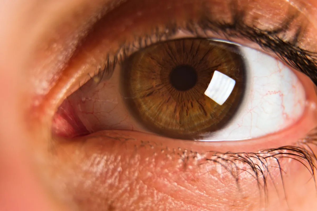
Inherited corneal disease, also known as hereditary corneal dystrophy, is a group of genetic disorders that affect the transparent front surface of the eye – the cornea. The cornea’s job is to let light in and keep the eye clear. In these disorders, abnormal protein or lipid deposits, scarring, or structural irregularities develop from birth or early puppyhood, leading to cloudiness, irritation, and sometimes ulceration.
There are several distinct forms, each linked to a different gene mutation. The most common are:
- Primary corneal dystrophy (PCD) – a simple, autosomal‑dominant condition causing bilateral, symmetric clouding.
- Granular corneal dystrophy (GCD) – characterized by granular, white‑specked lesions.
- Band keratopathy – calcium deposits that form a “band” across the cornea.
- Posterior polymorphous corneal dystrophy (PPCD) – affects the inner corneal layer and can lead to endothelial dysfunction.
According to the American College of Veterinary Ophthalmology (ACVO), hereditary corneal disease accounts for roughly 5–10 % of all corneal problems seen in dogs, making it a relatively common genetic eye issue.
What causes it?
Inherited corneal disease is caused by mutations in specific genes that control corneal structure and protein turnover. The mode of inheritance varies by type:
| Form | Gene (example) | Inheritance pattern |
|---|---|---|
| Primary corneal dystrophy | CHST6 (human analog) | Autosomal‑dominant |
| Granular corneal dystrophy | TGFBI | Autosomal‑dominant |
| Posterior polymorphous dystrophy | ZEB1 | Autosomal‑recessive or dominant (breed‑dependent) |
| Band keratopathy | Multiple metabolic genes | Complex, often multifactorial |
These mutations are passed from parent to offspring, meaning that if a breeding pair carries the gene, puppies have a predictable risk of inheriting the disease. Environmental factors (e.g., chronic irritation, vitamin A deficiency) can worsen the clinical picture, but the root cause remains genetic.
Signs and symptoms
Owners usually notice changes between 4 weeks and 2 years of age, though some forms present later in senior dogs. Early signs are subtle; they can progress to more serious complications if left unchecked.
| Severity | Typical signs |
|---|---|
| Mild | Hazy or slightly cloudy cornea, occasional blinking, mild photophobia (sensitivity to light). |
| Moderate | Visible white or gray speckles, recurrent tearing, occasional corneal ulceration, reduced shine in the eye. |
| Severe | Significant clouding, persistent ulcer, corneal neovascularization (new blood vessels), pain signs (pawing at eye, squinting), vision loss. |
Common complaints owners report include:
- “My dog keeps rubbing his eye on the carpet.”
- “She seems more hesitant to go outside on bright days.”
- “I noticed a gray ring around the iris when I looked closely.”
These symptoms can overlap with other ocular conditions, such as infectious ulcer or dry eye, so a professional exam is essential.
When to call your vet
Call your vet today if you notice:
- New cloudiness or speckling in either eye.
- Persistent tearing, redness, or discharge.
- Frequent blinking, squinting, or pawing at the eye.
- Any sign of pain (whining, reluctance to be touched).
Go to an emergency veterinary hospital right now if you see:
- A clear, bright red spot on the cornea (possible ulcer).
- Sudden vision loss or the dog is unable to navigate familiar surroundings.
- Excessive swelling, bleeding, or a large area of the cornea looks white and opaque.
These red‑flag signs can indicate an acute corneal ulcer or secondary infection that needs immediate treatment to prevent permanent vision loss.
This article is for informational purposes only and does not replace a hands‑on examination by your veterinarian.
How vets diagnose it
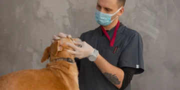
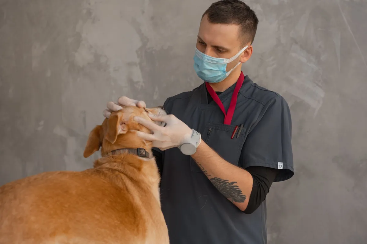
Diagnosis starts with a thorough eye exam. Your vet will use a slit‑lamp microscope to magnify the cornea, looking for characteristic deposits or scarring. A fluorescein stain is applied; the dye highlights any breaks in the corneal surface, turning bright green where an ulcer exists.
Additional tests may include:
- Schirmer tear test – measures tear production to rule out dry eye, which can worsen corneal disease.
- Intra‑ocular pressure (tonometry) – ensures the eye isn’t developing glaucoma.
- Corneal cytology or biopsy – rarely needed, but can confirm the type of dystrophy.
- Genetic testing – many labs now offer DNA panels for known corneal dystrophy genes. A buccal swab or blood sample can reveal whether a dog carries the mutation (source: UC Davis Veterinary Genetics Laboratory).
Imaging such as ocular ultrasound or anterior segment optical coherence tomography (AS‑OCT) may be used for deeper assessment, especially if a posterior dystrophy is suspected.
Treatment options
Medical treatment
Most hereditary corneal disease cases are managed medically, especially when the disease is mild to moderate. Common therapeutic classes include:
- Topical lubricants (artificial tears, hyaluronic acid) – keep the cornea moist and reduce friction.
- Anti‑inflammatory eye drops – corticosteroids (e.g., prednisolone acetate) or non‑steroidal anti‑inflammatory drugs (NSAIDs) to control inflammation. Ask your vet about this option.
- Antibiotic ointments – prevent secondary bacterial infection when an ulcer is present (e.g., neomycin‑polymyxin B).
- Vitamin A or omega‑3 supplements – may help maintain corneal health; discuss with your vet.
Systemic immunosuppressants (e.g., cyclosporine) are occasionally used for aggressive immune‑mediated forms, but they are reserved for cases that do not respond to topical therapy.
Supplements and supportive care
Evidence supports the use of omega‑3 fatty acids (EPA/DHA) from fish oil to reduce ocular surface inflammation. A daily dose of 20 mg EPA per kilogram of body weight is typical for dogs, but your vet will tailor the amount.
Probiotics (e.g., Enterococcus faecium) can support overall immune health, though they are not a direct treatment for corneal dystrophy. If your dog is on a dry‑food diet, adding a modest amount of canned pumpkin or low‑fat yogurt can improve hydration, which may benefit tear film quality.
Procedures or surgery
When the cornea becomes ulcerated or the clouding threatens vision, surgical intervention may be required:
- Corneal debridement – gentle removal of the diseased superficial layer to promote healing.
- Phototherapeutic keratectomy (PTK) – laser resurfacing that removes deposits and smooths the corneal surface.
- Penetrating keratoplasty (corneal transplant) – replacement of the full‑thickness cornea with donor tissue. This is a specialty procedure, usually performed at a veterinary ophthalmology referral center.
- Therapeutic contact lenses – protect the cornea while it heals after surgery.
Post‑operative care often includes intensive topical medication, protective eye patches, and close monitoring for infection. The success rate for PTK in hereditary dystrophy is about 70–80 % according to recent ACVO data, while full corneal transplants have a higher complication risk but can restore vision in many cases.
Diet and nutrition
While no diet can cure a genetic corneal defect, proper nutrition can support overall eye health, reduce inflammation, and improve tear film quality. Here are practical feeding guidelines for dogs with hereditary corneal disease:
| Do feed | Limit | Avoid |
|---|---|---|
| High‑quality protein sources (chicken, turkey, fish) that are highly digestible. | Excessive calcium supplements – can promote mineral deposits. | High‑fat, low‑moisture kibble that may decrease overall hydration. |
| Omega‑3 rich foods (salmon, sardines, fish oil) or a veterinary‑approved omega‑3 supplement. | Artificial colors and flavors – little benefit for eye health. | Excessive vitamin A from liver treats – can lead to hypervitaminosis A. |
| Prescription “ocular health” diets (e.g., renal‑support or low‑protein therapeutic foods) if the dog also has kidney issues, as recommended by your vet. | High‑salt snacks – may increase intra‑ocular pressure. | Raw diets with unbalanced calcium‑phosphorus ratios unless formulated by a veterinary nutritionist. |
Key points to remember:
- Provide fresh water at all times; a humidifier in dry climates can help keep the ocular surface moist.
- Consider adding a small amount of canned pumpkin or unsweetened yogurt to the daily meal for extra moisture and beneficial probiotics.
- If your dog is on a therapeutic eye diet (e.g., those formulated for keratoconjunctivitis sicca), follow the feeding schedule your vet prescribes and transition slowly over 7–10 days to avoid gastrointestinal upset.
- Monitor weight – obesity can worsen inflammation and complicate surgical recovery.
For a personalized feeding plan, use our dog nutrition calculator or consult a board‑certified veterinary nutritionist.
Cost and prognosis
Financial considerations are a real part of managing hereditary corneal disease. Below are typical cost ranges (estimates, may vary by region and clinic):
| Service | US (USD) | UK (GBP) |
|---|---|---|
| Initial ophthalmic exam + fluorescein staining | $120–$200 | £90–£150 |
| Genetic test panel (single gene or panel) | $150–$300 | £110–£250 |
| Topical medication (3‑month supply) | $80–$200 | £60–£150 |
| Phototherapeutic keratectomy (PTK) | $1,500–$3,000 | £1,200–£2,400 |
| Full‑thickness corneal transplant | $4,000–$7,500 | £3,200–£5,800 |
Prognosis depends on the specific dystrophy, age at diagnosis, and whether complications such as ulceration or glaucoma develop. In many dogs, especially those with primary corneal dystrophy, vision can be preserved for years with diligent medical care. Surgical options like PTK can improve clarity and comfort in moderate cases. Full corneal transplants carry a higher risk of rejection (about 20 % in dogs) but can restore near‑normal vision when successful.
Overall quality‑of‑life is good for most dogs that receive timely treatment. Senior dogs may experience slower healing, so realistic expectations and close follow‑up are essential.
Prevention and home care
Because the root cause is genetic, true prevention requires responsible breeding practices. Here’s what owners and breeders can do:
- Genetic screening – Have breeding dogs tested for known corneal dystrophy mutations. Only breed dogs that test clear or are carriers with a non‑carrier partner.
- Regular eye exams – Schedule a veterinary ophthalmology check at 6 months, then annually. Early detection catches subtle clouding before it progresses.
- Protect the eyes – Avoid exposure to harsh chemicals, smoke, or prolonged UV light. Sunglasses for dogs (e.g., Doggles) can reduce UV‑induced damage in high‑risk breeds.
- Maintain tear film – Use preservative‑free artificial tears if the dog shows chronic dryness. A humidifier in dry indoor environments can help.
- Monitor for trauma – Keep nails trimmed and discourage rough play that could scratch the cornea.
For owners of dogs already diagnosed with hereditary corneal disease, a daily routine of gentle eye cleaning with a sterile saline wipe can prevent debris buildup and infection. Keep a log of any changes in cloudiness, discharge, or behavior, and share it with your vet at each visit.
From our vet team: “If you catch a cloudy cornea early, most dogs do fine with just drops and a little extra care. The biggest mistake owners make is waiting until the eye hurts before seeking help – early intervention is the key to preserving vision.”
Key takeaways
- Inherited corneal disease is a genetic eye disorder that causes clouding, scarring, or ulceration, most often seen in specific breeds.
- Early signs include hazy eyes, mild tearing, and increased light sensitivity; any sudden pain or ulcer warrants immediate veterinary attention.
- Diagnosis relies on a slit‑lamp exam, fluorescein staining, and increasingly on DNA testing to confirm the specific mutation.
- Medical management (lubricants, anti‑inflammatories, antibiotics) works for many dogs; surgical options like PTK or corneal transplant are reserved for advanced cases.
- Feeding a high‑quality, omega‑3‑rich diet and maintaining proper hydration supports ocular health, but no diet can reverse the genetic defect.
- Responsible breeding, regular eye exams, and prompt treatment together give dogs the best chance at a comfortable, vision‑preserving life.
Myth vs. fact
Myth: All hereditary corneal disease leads to blindness.
Fact: Most dogs retain functional vision for years, especially when the disease is caught early and managed with medication or laser therapy.
Myth: A cloudy cornea always means a painful ulcer.
Fact: Cloudiness can be a benign dystrophy without pain; however, any sudden increase in opacity or a bright green fluorescein stain signals an ulcer that needs urgent care.
Myth: Genetic testing is too expensive and not worth it.
Fact: DNA panels for corneal dystrophy are now under $300 and can prevent the spread of the trait in breeding programs, saving money and heartache long‑term.
Frequently asked questions
What causes inherited corneal disease in dogs?
It’s caused by specific gene mutations that affect corneal protein or lipid metabolism, passed from parent to offspring in autosomal‑dominant or recessive patterns.
How can I tell if my dog has a hereditary corneal problem?
The first clue is a hazy or speckled cornea, often noticed in puppies. A vet can confirm it with a slit‑lamp exam, fluorescein stain, and optionally a DNA test.
Are there any cures for genetic corneal disease in dogs?
There’s no cure that changes the underlying DNA, but medical treatments can control symptoms, and laser or transplant surgeries can restore clarity and vision in many cases.
Will my dog lose vision from inherited corneal disease?
Most dogs maintain usable vision for years, especially with early treatment. Vision loss is more likely if ulcers, infections, or glaucoma develop and aren’t treated promptly.
Is genetic testing available for corneal disorders in dogs?
Yes. Several veterinary labs, including UC Davis and commercial providers, offer DNA panels that identify the common corneal dystrophy mutations.
What is the typical cost of treating hereditary corneal disease?
Initial exam and testing range from $120–$300; medication adds $80–$200 per three months; laser (PTK) costs $1,500–$3,000; full corneal transplant can exceed $4,000 in the United States.
Ask the PuppaDogs community
Have a question this article didn’t fully answer? Want to compare notes with other dog owners who’ve been through this? Our community forum is moderated by experienced owners and vets — and answers tend to come fast. Ask in the PuppaDogs community →
References
- American College of Veterinary Ophthalmology (ACVO). “Corneal Dystrophies in Dogs.” 2023.
- American Animal Hospital Association (AAHA). “Guidelines for Ophthalmic Care.” 2022.
- Merck Veterinary Manual. “Corneal Disease, Hereditary.” Updated 2024.
- Cornell University College of Veterinary Medicine. “Inherited Corneal Dystrophies.” 2023.
- UC Davis Veterinary Genetics Laboratory. “DNA Testing for Canine Corneal Dystrophy.” 2022.
- World Small Animal Veterinary Association (WSAVA). “Ocular Health Recommendations.” 2022.
- American Veterinary Medical Association (AVMA). “Pain Management in Veterinary Ophthalmology.” 2021.
- International Veterinary Ophthalmology Society. “Phototherapeutic Keratectomy Outcomes.” 2023.


