


Quick take: Eosinophilic meningoencephalomyelitis (EME) is a rare, immune‑mediated inflammation of the brain and spinal cord that can cause seizures, weakness, and pain. Prompt veterinary care, targeted immunosuppressive drugs, and supportive care give many dogs a good chance at recovery, but the disease can be life‑threatening if left untreated.
It’s 11 p.m., and your normally boisterous Border Collie is lying still on the couch, eyes half‑closed, and you notice his gums look a little pink‑ish instead of the usual bright red. You scroll through a late‑night search and the term “eosinophilic meningoencephalomyelitis” pops up. Your heart races—could this be something serious? You’re not alone; many owners first spot the subtle change in energy or gait before the diagnosis lands.
In this article we’ll explain exactly what brain and spinal cord inflammation (eosinophilic meningoencephalomyelitis) is, why it happens, what signs to watch for, and how vets diagnose and treat it. We’ll also cover nutrition, costs, prognosis, and steps you can take at home to help your dog feel better faster. By the end you’ll have a clear roadmap for the next vet visit and a realistic view of what recovery looks like.
What is brain and spinal cord inflammation (eosinophilic meningoencephalomyelitis) in dogs?
Eosinophilic meningoencephalomyelitis (EME) is an inflammatory disease that targets the brain (meningo‑encephalitis) and spinal cord (myelitis) simultaneously. “Eosinophilic” refers to a type of white blood cell—eosinophils—that pile up in the central nervous system (CNS) and release chemicals that cause swelling, pain, and tissue damage.
In healthy dogs, the immune system protects against infections. In EME, the immune system mistakenly attacks the CNS, a process called autoimmune inflammation. The exact trigger is still unclear, but the condition is most commonly seen in young to middle‑aged dogs, especially certain breeds such as the Golden Retriever, Beagle, and German Shepherd.
Although EME is considered rare—accounting for less than 1 % of all canine neurologic cases—it is well‑documented in veterinary neurology literature and recognized by the American College of Veterinary Internal Medicine (ACVIM). Early recognition and treatment are crucial because inflammation can quickly progress to irreversible nerve damage.
What causes it?
The precise cause of eosinophilic meningoencephalomyelitis remains unknown, but several risk factors and possible triggers have been identified:
| Category | Potential contributors |
|---|---|
| Genetic predisposition | Breed‑specific susceptibility (e.g., Golden Retrievers, Beagles, German Shepherds) noted in ACVIM surveys. |
| Immune dysregulation | Autoimmune mechanisms where the body mistakenly targets CNS tissue; parallels with human eosinophilic meningitis. |
| Environmental triggers | Exposure to certain parasites (e.g., Angiostrongylus spp.) or allergens that may stimulate eosinophil activity, though direct causation is unproven. |
Because the disease is immune‑mediated, many vets treat it similarly to other autoimmune neurologic disorders, using immunosuppressive drugs to calm the misguided immune response.
Signs and symptoms
EME can affect both the brain and spinal cord, so the clinical picture varies widely. Early signs are often subtle, while later stages can become severe. Below is a typical progression:
| Stage | Common signs |
|---|---|
| Mild | Reduced playfulness, slight wobble on walks, occasional head tilt, mild facial twitching. |
| Moderate | Seizures (generalized or focal), noticeable weakness in one or more limbs, loss of coordination (ataxia), persistent head tilt, decreased appetite. |
| Severe | Paralysis, severe pain (crying or yelping when moving), stupor or coma, rapid breathing, vomiting, and signs of increased intracranial pressure such as bulging eyes. |
Other red‑flag clues include pale or bluish gums, fever, and a sudden change in behavior (e.g., aggression or confusion). Because eosinophils can also accumulate in other organs, some dogs develop respiratory signs or skin itching, but the neurologic symptoms dominate the clinical picture.
When to call your vet
Call your vet today if you notice any of the following:
- New or worsening seizures.
- Sudden weakness or loss of coordination.
- Persistent head tilt or facial twitching.
- Pale, bluish, or mottled gums.
- Refusal to eat or drink for more than 12 hours.
Go to an emergency veterinary hospital right now if you see:
- Complete paralysis of one or more limbs.
- Severe pain with vocalization when the dog moves.
- Signs of brain swelling: rapid breathing, vomiting, or a dilated pupil.
- Unconsciousness or collapse.
These signs can indicate a medical emergency that requires immediate stabilization. Remember, this article is for information only; it does not replace a hands‑on exam by a qualified veterinarian.
How vets diagnose it
Diagnosing eosinophilic meningoencephalomyelitis involves a stepwise approach:
- History and physical exam: The vet will ask about the onset of neurologic signs, diet, travel, and any recent illnesses. A thorough neurologic exam pinpoints which parts of the CNS are affected.
- Blood work: A complete blood count (CBC) often reveals eosinophilia (high eosinophil count) and may show inflammatory markers. Biochemistry panels assess organ function before starting steroids.
- Cerebrospinal fluid (CSF) analysis: A sample taken via lumbar puncture can show increased eosinophils, elevated protein, and a characteristic “pleocytosis.” CSF cytology is key for confirming an eosinophilic component.
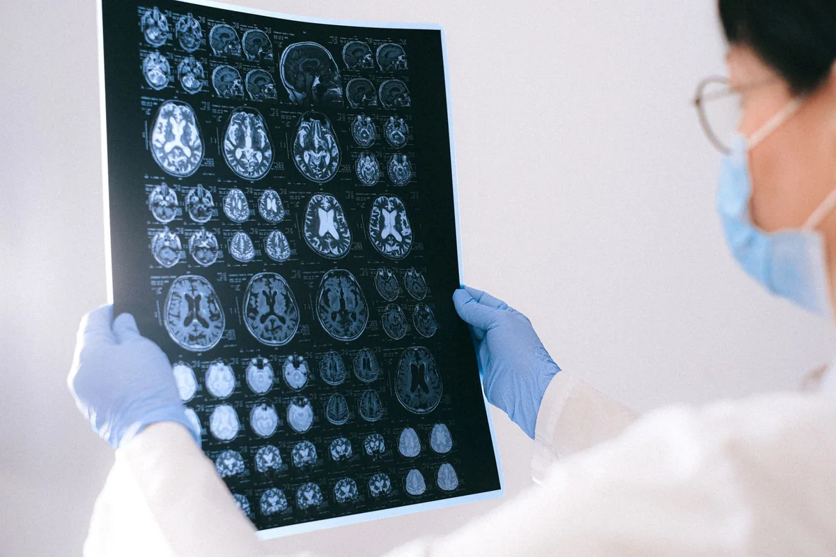
- Advanced imaging: MRI of the brain and spinal cord highlights areas of inflammation, edema, and any lesions. Contrast‑enhanced MRI is the gold standard for visualizing active inflammation.
- Rule out infections: Tests for parasites (e.g., Angiostrongylus), bacterial cultures, and viral PCRs are performed to exclude infectious meningitis, which requires different treatment.
Only after infectious causes are excluded and CSF shows eosinophilic inflammation will a diagnosis of EME be made, according to ACVIM guidelines.

Treatment options
Medical treatment
Immunosuppression is the cornerstone of therapy. Common drug classes include:
- Corticosteroids: Prednisone or dexamethasone are usually started at a high dose to rapidly dampen inflammation. Your vet will tailor the dose to your dog’s weight and severity.
- Additional immunosuppressants: Drugs such as azathioprine, mycophenolate mofetil, or cyclosporine may be added to reduce steroid dependence and control chronic inflammation.
- Anticonvulsants: If seizures are present, medications like levetiracetam or phenobarbital are prescribed to stabilize brain activity.
- Supportive anti‑inflammatory agents: NSAIDs are generally avoided during high‑dose steroid therapy because of gastrointestinal risk, but low‑dose aspirin may be used under vet supervision.
All medications should be discussed with your vet; ask about potential side effects and the expected tapering schedule.
Supplements and supportive care
While no supplement can replace immunosuppressive drugs, certain adjuncts can aid recovery:
- Omega‑3 fatty acids (EPA/DHA): These have anti‑inflammatory properties and may help reduce CNS swelling. A high‑quality fish‑oil supplement (e.g., 1,000 mg EPA/DHA per 10 kg body weight) is often recommended.
- Antioxidants: Vitamin E and selenium support neuronal health, especially during steroid therapy which can increase oxidative stress.
- Probiotics: Maintaining gut health can improve overall immunity and aid in the absorption of oral medications.
Supplements should be introduced gradually and only after consulting your vet, as they can interact with immunosuppressants.
Procedures or surgery
In most cases EME is managed medically. However, if MRI reveals a focal mass effect or severe spinal cord compression, a neurosurgical decompression may be considered. This is rare and typically performed at specialty referral centers. Recovery from CNS surgery can take 4–6 weeks, and costs often range from $5,000 to $12,000 USD (or equivalent in the UK), depending on the complexity and hospital fees.
Diet and nutrition
Nutrition plays a supportive role in managing eosinophilic meningoencephalomyelitis. While there is no “cure‑diet,” feeding strategies that reduce inflammation and support healing can improve your dog’s overall resilience.
Key principles:
- Highly digestible protein: Choose a diet with quality animal protein (e.g., chicken, turkey, or fish) that is easy on the gastrointestinal tract. This helps maintain muscle mass while the dog recovers.
- Moderate fat, high omega‑3: Adding a fish‑oil supplement boosts EPA/DHA, which have documented anti‑inflammatory effects on the CNS. Look for commercial foods labeled “with added omega‑3” or supplement separately.
- Low‑carbohydrate, limited simple sugars: Reducing excess glucose can prevent secondary inflammation. Grain‑free or limited‑grain formulas often meet this goal.
- Antioxidant‑rich ingredients: Foods containing blueberries, spinach, or added vitamin E can help counter oxidative stress from steroid therapy.
- Limited sodium: High sodium can exacerbate cerebral edema. Choose diets with < 0.3 % sodium on a dry‑matter basis.
Many owners find that a therapeutic “neurology support” or “anti‑inflammatory” diet—such as those formulated for dogs with epilepsy or neurodegenerative disease—meets these criteria. These diets are usually prescription‑only; discuss options with your vet. If you prefer home‑cooked meals, a balanced recipe might include boiled chicken breast, steamed pumpkin, a teaspoon of fish oil, and a sprinkle of blueberries.
Below is a quick reference for feeding choices:
| Do feed | Limit | Avoid |
|---|---|---|
| Lean animal protein (chicken, turkey, fish) | Cooked starchy carbs (sweet potato, rice) – keep portions moderate | High‑fat treats, fried foods, processed snacks |
| Omega‑3 sources (fish oil, salmon) | Commercial grain‑free kibble with unknown fillers | Excessive sodium (cured meats, cheese) |
| Antioxidant foods (blueberries, spinach) | High‑sugar treats (cookies, fruit juices) | Raw bones that could splinter |
When transitioning to a new diet, do it gradually over 7–10 days: mix 25 % new food with 75 % old, then increase the new portion each few days. This helps avoid GI upset, which can be problematic when your dog is already on steroids.
Hydration is also essential. Steroids can increase thirst and urination, so ensure fresh water is always available. Some owners add a low‑sodium broth to meals to encourage fluid intake without adding excess salt.
Finally, coordinate diet changes with your vet’s medication plan. Certain foods can affect drug absorption (e.g., high‑fat meals may delay oral steroid uptake). Your vet may suggest feeding the medication with a small amount of food rather than on an empty stomach.
Cost and prognosis
Because EME is a specialist-level neurologic disease, costs can vary widely. Below are typical ranges (estimated, based on US and UK veterinary pricing):
| Item | US estimate | UK estimate |
|---|---|---|
| Initial consult + MRI | $1,200–$2,500 | £800–£1,500 |
| CSF analysis + lab work | $300–$600 | £150–£300 |
| Immunosuppressive drugs (first 3 months) | $200–$600 | £120–£350 |
| Follow‑up visits (3‑month period) | $150–$300 | £80–£200 |
| Potential surgery (rare) | $5,000–$12,000 | £4,000–£9,000 |
Overall, owners can expect to spend roughly $2,000–$4,000 USD (or £1,200–£2,500) for the first six months of care, not including emergency interventions.
Prognosis depends on how quickly treatment starts and the severity of neurologic signs. Studies published by the American College of Veterinary Internal Medicine suggest:
- ~60–70 % of dogs achieve partial or full neurologic recovery if treatment begins within two weeks of symptom onset.
- ~30 % may have persistent deficits (e.g., mild weakness or ataxia) despite therapy.
- ~10 % may not survive the acute phase, especially if severe brain swelling or paralysis is present.
Long‑term quality of life is generally good for dogs that respond to therapy, though many require lifelong low‑dose immunosuppression and periodic neurologic exams.
Prevention and home care
Because the exact trigger for EME is unknown, true prevention is challenging. However, you can reduce risk and support recovery by:
- Maintaining a balanced diet: A diet rich in omega‑3 fatty acids and antioxidants can help keep the immune system in check.
- Regular parasite control: Use year‑round heartworm and intestinal parasite preventatives (consult your vet for region‑appropriate products; see our dog‑health community for recommendations).
- Minimizing exposure to known allergens: If your dog has a history of skin allergies, keep the home environment clean and avoid known irritants.
- Routine wellness exams: Annual or semi‑annual check‑ups can catch early neurologic changes before they become severe.
- Monitoring behavior: Keep a daily log of activity, appetite, and any subtle gait changes. Early detection improves treatment success.
- Stress reduction: Limit sudden environmental changes (new pets, moving houses) that can exacerbate immune dysregulation.
If your dog has already been diagnosed, follow-up appointments every 4–6 weeks during the tapering phase are typical. Your vet may use a weight‑based dosage calculator to adjust medication doses precisely.
From our vet team: “Most owners worry that an ‘eosinophilic’ label means a parasite is the culprit. In reality, it signals an immune response, and the treatment focus is on calming that response. Prompt, aggressive immunosuppression combined with supportive nutrition gives many dogs a solid chance at regaining normal function.”
Key takeaways
- Eosinophilic meningoencephalomyelitis is a rare, immune‑mediated inflammation of the brain and spinal cord that can cause seizures, weakness, and pain.
- Early signs include subtle gait changes, head tilt, and reduced energy; severe signs require immediate veterinary or emergency care.
- Diagnosis relies on blood work, CSF analysis, and MRI to rule out infections and confirm eosinophilic inflammation.
- Treatment centers on high‑dose steroids and additional immunosuppressants, with omega‑3 supplements often added for anti‑inflammatory support.
- Feeding a high‑quality, digestible protein diet enriched with omega‑3s and antioxidants aids recovery and helps manage steroid side effects.
- Prognosis is good for dogs treated early, but costs can range from a few thousand dollars to over ten thousand if surgery is needed.
Myth vs. fact
Myth: Eosinophilic meningoencephalomyelitis is caused by a worm infection.
Fact: While parasites can raise eosinophil levels, EME is primarily an autoimmune condition; parasites are ruled out before confirming the diagnosis.
Myth: Once a dog starts on steroids, it will be on them for life.
Fact: Most dogs taper off high‑dose steroids within months, transitioning to a low maintenance dose or alternative immunosuppressants under veterinary guidance.
Myth: Diet has no impact on neurologic diseases.
Fact: Proper nutrition, especially omega‑3 fatty acids and antioxidants, can reduce inflammation and support recovery during immunosuppressive therapy.
Frequently asked questions
What is the typical cost of diagnosing and treating EME?
Initial diagnostics (consult, MRI, CSF analysis) usually cost $1,500–$3,000 USD, and the first three months of medication add $200–$600. Surgery, if required, can push the total beyond $10,000.
Is eosinophilic meningoencephalomyelitis contagious?
No. EME is an autoimmune condition, not an infectious disease, so it cannot be passed between dogs or to humans.
How long does recovery take after treatment begins?
Most dogs show noticeable improvement within 2–4 weeks of aggressive immunosuppression. Full neurologic recovery can take 3–6 months, with periodic rechecks to monitor progress.
Can my dog live a normal life after EME?
Yes, many dogs return to their pre‑illness activity levels after treatment and a careful tapering schedule. Some may have mild residual weakness, but quality of life is generally high.
Are there any home remedies that can replace veterinary medication?
Supplements like fish oil can support anti‑inflammatory processes, but they are not a substitute for prescription immunosuppressants. Always discuss any additions with your vet.
What should I ask my vet at the next appointment?
Ask about the specific immunosuppressive protocol, expected side effects, how to monitor for relapse, and whether a therapeutic diet is recommended for your dog’s breed and condition.
Ask the PuppaDogs community
Have a question this article didn’t fully answer? Want to compare notes with other dog owners who’ve been through this? Our community forum is moderated by experienced owners and vets — and answers tend to come fast. Ask in the PuppaDogs community →
References
- American College of Veterinary Internal Medicine (ACVIM). “Autoimmune Neurologic Diseases in Dogs” – 2023 guidelines.
- Merck Veterinary Manual. “Meningoencephalomyelitis, Eosinophilic” – Chapter on CNS inflammatory disorders.
- American Veterinary Medical Association (AVMA). “Steroid Use in Veterinary Medicine” – 2022 update.
- World Small Animal Veterinary Association (WSAVA). “Nutrition for Dogs with Neurologic Disease” – 2021 position statement.
- AAHA. “Diagnostic Imaging in Neurologic Cases” – 2022 recommendations.
- Cornell University College of Veterinary Medicine. “CSF Analysis Interpretation” – 2020 reference.
- Plumb’s Veterinary Drug Handbook. “Immunosuppressive Agents in Dogs” – 9th edition, 2022.
- American Animal Hospital Association (AAHA). “Pain Management in Dogs” – 2023 guidelines.
