


Quick take: Blood in the chest cavity—called a hemothorax—is usually caused by trauma, a tumor, or a bleeding disorder. It can be life‑threatening, but prompt veterinary care (often a chest tube or surgery) gives many dogs a good chance of recovery. Call your vet right away if you notice rapid breathing, pale gums, or a sudden collapse.
It’s 11 p.m., the hallway lights are on, and your two‑year‑old mixed‑breed Labrador isn’t greeting you at the door. Instead, he lies on the floor, breathing shallowly, and when you gently lift his head you see his gums are a stark, almost gray‑white. Your heart races, you Google “blood in the chest in dogs,” and the search results start flashing medical terms you barely understand.
We get it—seeing your dog in distress is terrifying, and the internet can make the situation feel even more urgent. The good news is that “blood in the chest” has a specific name (hemothorax), and while it can be serious, there are clear steps you can take right now and a roadmap for veterinary treatment. In this article we’ll explain what a hemothorax is, why it happens, what signs to watch for, how vets diagnose it, the treatment options (including costs), and how you can help your dog recover at home.
What is hemothorax?
Hemothorax (pronounced hee‑MO‑thor‑aks) is the medical term for blood that accumulates in the thoracic cavity—the space between the lungs and the chest wall. In a healthy dog, this space contains a thin layer of fluid that lets the lungs expand and contract smoothly. When blood fills the cavity, it compresses the lungs, limits oxygen exchange, and can quickly become a life‑threatening emergency.
Hemothorax is relatively uncommon compared with other chest problems like pneumothorax (air in the chest). Large‑breed dogs and older dogs are seen more often, but any dog can develop it if the underlying cause is present. According to the American College of Veterinary Internal Medicine (ACVIM), traumatic hemothorax accounts for roughly 60 % of cases, while spontaneous (non‑traumatic) bleeding makes up the remainder.
What causes it?
Blood can enter the chest cavity for several reasons. The most frequent categories are trauma, tumors, and clotting disorders. Below is a quick reference to help you identify the likely cause based on your dog’s history.
| Cause | Typical scenario | Notes |
|---|---|---|
| Trauma | Car accident, fall from height, blunt impact | Often accompanied by rib fractures or internal organ injury. |
| Neoplasia (tumor) | Chest wall or lung tumor that erodes vessels | More common in middle‑aged to senior dogs; breeds like German Shepherds and Boxers have higher incidence. |
| Coagulopathy | Bleeding disorders (e.g., hemophilia, rodenticide poisoning, severe liver disease) | May be spontaneous; blood tests reveal clotting abnormalities. |
| Ruptured blood vessels | Large vessel aneurysm, severe hypertension, or iatrogenic injury during procedures | Rare but can cause sudden, massive bleeding. |
| Other | Parasites (heartworm), severe infections, or autoimmune vasculitis | These are less common and usually diagnosed alongside other systemic signs. |
Signs and symptoms
Because the chest cavity is a closed space, even a modest amount of blood can cause noticeable problems. Early signs are often subtle, while severe cases present as an emergency.
| Severity | Typical signs |
|---|---|
| Mild | Reduced stamina on walks, slight cough, occasional panting, faint pink‑to‑pale gums. |
| Moderate | Labored breathing, rapid shallow breaths, persistent cough, reluctance to lie down, noticeable abdominal effort. |
| Severe | Sudden collapse, very pale or gray gums, bluish tongue, inability to rise, shock (weak pulse, cold extremities). |
Other red‑flag clues include a “gurgling” sound when you listen to the chest with a stethoscope, a visible bulge in the side of the ribcage, or an obvious wound that could be bleeding into the chest.
When to call your vet
Call your vet today if you notice any of the following:
- Persistent cough or gagging that’s new for your dog.
- Noticeably pale, gray, or bluish gums.
- Rapid, shallow breathing or difficulty getting a full breath.
- Sudden weakness, reluctance to move, or stumbling.
- Any recent trauma (car hit, fall, rough play) even if there’s no obvious wound.
Go to an emergency veterinary hospital right now if your dog shows any sign of shock:
- Collapse or inability to stand.
- Very pale or white gums, tongue, or inside of the eyelids.
- Very fast heart rate (more than 150 bpm) or weak pulse.
- Cold front paws or ears.
This article is for informational purposes only and does not replace professional veterinary care. Always seek immediate veterinary attention if you suspect a hemothorax.
How vets diagnose hemothorax
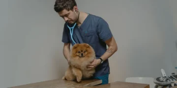
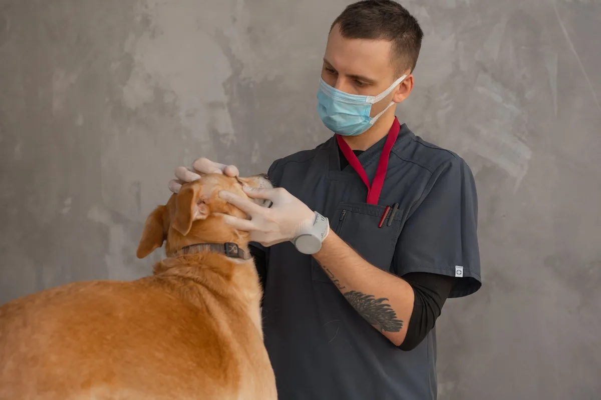
Diagnosis starts with a thorough history and physical exam. Your vet will listen to the chest, check gum color, and feel for any rib abnormalities.
- Thoracic radiographs (X‑rays): Blood appears as a dense, fluid‑filled area that settles in the dependent (lowest) part of the chest. The “fluid line” can be distinguished from air on the film.
- Ultrasound (Thoracic ultrasound): Provides a real‑time view of fluid accumulation and can guide needle placement for fluid removal.
- Computed tomography (CT scan): Offers detailed images of the chest structures and helps locate the source of bleeding when X‑rays are inconclusive.
- Fluid analysis: If fluid is tapped, it is examined for red blood cell count, clotting factors, and possible infection.
- Blood work: Complete blood count (CBC) and coagulation profile assess anemia and clotting status; a low hematocrit supports ongoing bleeding.
These diagnostics let the veterinary team determine how much blood is present, whether the bleeding is ongoing, and what underlying cause to target.
Treatment options
Medical treatment
When the bleeding source is controllable or the dog is stable, medical management may be enough. Common drug classes include:
- Intravenous fluids: To maintain blood pressure and support circulation.
- Blood transfusions: If the dog is anemic, packed red blood cells restore oxygen‑carrying capacity.
- Immunosuppressants (e.g., prednisolone, cyclosporine): Used when a clotting disorder or immune‑mediated disease is suspected. Ask your vet about these options.
- Antibiotics: If a bacterial infection is present or the chest has been penetrated by a wound.
- Vitamin K or plasma: For rodenticide poisoning or other clotting factor deficiencies.
Supplements and supportive care
While supplements cannot stop active bleeding, they can aid recovery once the blood is removed and the lungs re‑expand.
- Omega‑3 fatty acids (EPA/DHA): Anti‑inflammatory properties that may help reduce scar tissue in the pleura.
- Probiotics: Support gut health after antibiotics or stress.
- Vitamin C: Antioxidant that some clinicians use to support vascular health, though evidence is limited.
Always discuss supplement use with your vet, especially if your dog is on blood thinners or other medications.
Procedures or surgery
Most hemothoraces require active drainage. The two main procedures are:
- Thoracocentesis: A needle is inserted into the chest to aspirate blood. This is often the first step to relieve pressure and confirm the diagnosis.
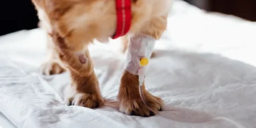
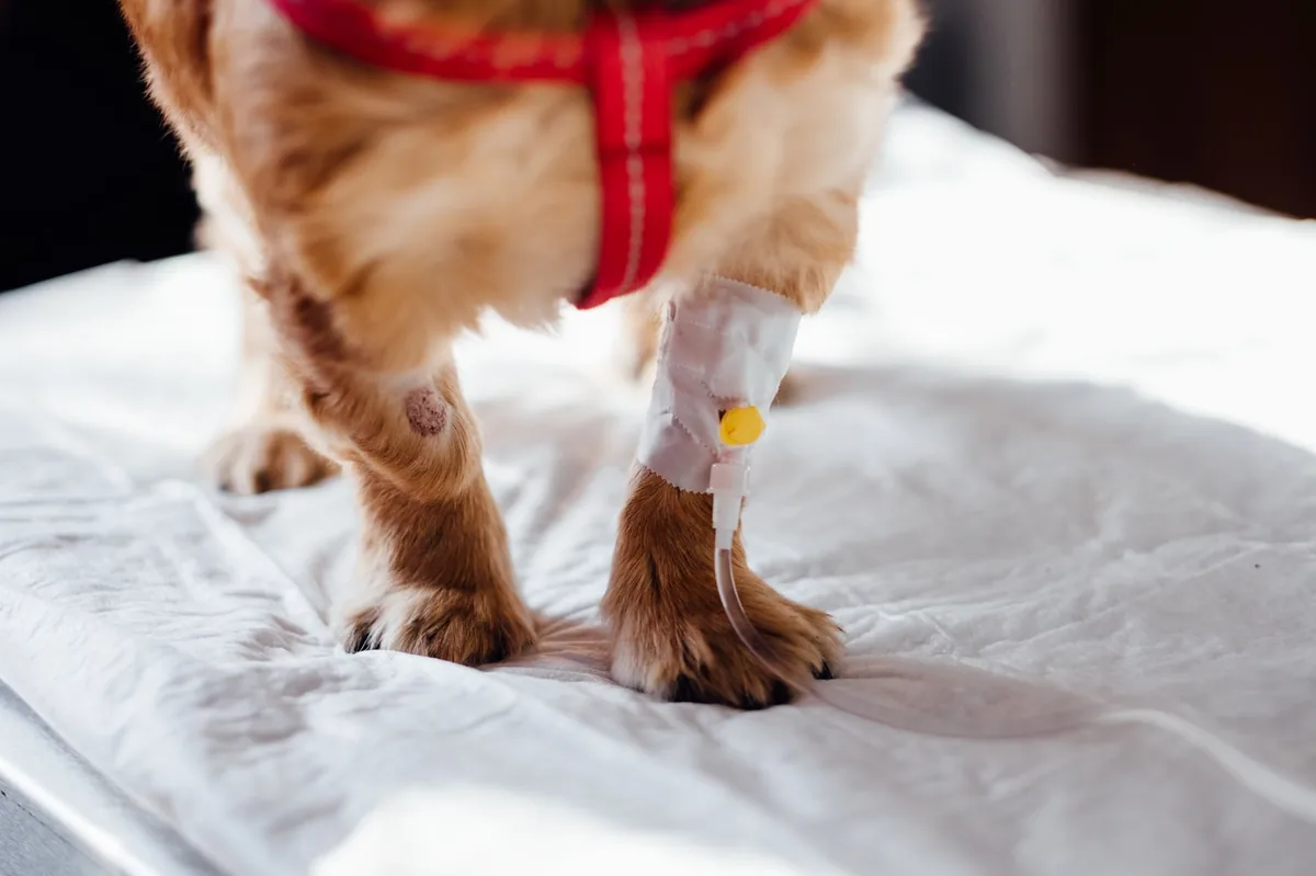
- Chest tube placement (thoracostomy tube): A small tube remains in the chest for 24–72 hours to continuously evacuate blood and prevent re‑accumulation. The tube is connected to a suction device and monitored closely.
- Surgical intervention: If a tumor, ruptured vessel, or severe organ damage is identified, thoracic surgery may be needed to repair the source. This is performed by a board‑certified veterinary surgeon and usually involves a thoracotomy (opening the chest wall).
Recovery from chest tube placement typically involves pain management, limited activity, and frequent re‑checks with X‑rays to ensure the space stays clear.
Diet and nutrition
Nutrition plays a crucial role in healing after a hemothorax, especially if surgery or a blood transfusion was required. The goals are to support blood production, maintain a healthy weight, and avoid foods that could worsen inflammation.
Protein: High‑quality, highly digestible protein helps rebuild red blood cells and repair tissue. Look for foods that list a named animal protein (chicken, salmon, lamb) as the first ingredient. If your dog is recovering from surgery, a protein content of 22‑28 % of the diet is ideal (AAFCO 2022).
Fat: Moderate fat (10‑15 % of calories) provides energy without overloading the liver. Omega‑3 sources such as fish oil can reduce inflammation in the pleural lining.
Carbohydrates: Easily digestible carbs (like rice or sweet potato) are fine, but avoid excessive fillers that can cause gastrointestinal upset.
Vitamins and minerals: Iron‑rich foods (e.g., cooked lean beef, organ meats) support red blood cell production, while vitamin B12 and folic acid are essential for marrow health. However, supplementing iron without veterinary guidance can be harmful, so ask your vet before adding any iron‑rich treats.
Many owners wonder whether a prescription “renal” or “post‑operative” diet is needed. In most hemothorax cases, a standard high‑protein diet is sufficient, unless your dog has a concurrent kidney disease. If a specific therapeutic diet is recommended, your vet will choose a brand that meets AAFCO nutrient profiles without marketing bias.
Below is a quick reference for feeding during recovery:
| Food category | Do feed | Limit | Avoid |
|---|---|---|---|
| High‑quality kibble | Yes – choose one with ≥22 % protein | Very high‑fat kibble | Low‑protein, filler‑heavy diets |
| Cooked home‑made meals | Yes – lean meat + rice/sweet potato + veg | Excessive butter or oil | Raw bones (risk of choking) |
| Supplements | Omega‑3 oil (vet approved) and probiotic | Iron tablets without vet guidance | Vitamin D megadoses |
| Treats | Lean jerky strips, small amounts of cheese | High‑salt snacks | Chocolate, grapes, onions |
When transitioning back to normal feeding, do it gradually over 3–5 days to avoid gastrointestinal upset. Offer smaller, more frequent meals (e.g., 3–4 times daily) and monitor stool consistency. Hydration is also key; fresh water should always be available, and you can add a splash of low‑sodium broth to encourage drinking.
Cost and prognosis
Financial considerations are an important part of emergency care. Below are typical cost ranges (as of 2024) for the United States and United Kingdom; actual fees vary by clinic, region, and severity.
| Service | US (USD) | UK (GBP) |
|---|---|---|
| Emergency exam & initial X‑ray | $150–$300 | £120–£250 |
| Thoracocentesis (single needle aspiration) | $200–$400 | £150–£300 |
| Chest tube placement (including monitoring 24–72 h) | $800–$2,200 | £700–£1,800 |
| Blood transfusion (one unit) | $250–$500 | £200–£400 |
| Thoracic surgery (if needed) | $3,000–$6,500 | £2,500–£5,500 |
| Post‑operative meds & follow‑up X‑rays | $200–$600 | £150–£450 |
Prognosis depends on the cause, amount of blood, and how quickly treatment begins. Dogs with traumatic hemothorax that receive prompt drainage have survival rates of 70‑85 % according to a 2022 ACVIM review. Spontaneous bleeding from tumors or severe clotting disorders carries a more guarded outlook, with survival ranging from 30‑60 %.
Long‑term quality of life is generally good if the underlying cause is resolved and the lungs re‑expand fully. Some dogs may develop mild pleural scarring, which can cause a slight decrease in stamina, but most return to normal activity within 4–6 weeks.
Prevention and home care
While you can’t always prevent an accident, several steps can reduce the risk of a hemothorax or aid early detection:
- Safe environments: Use leash walks in high‑traffic areas, secure fences, and avoid leaving toys that could cause a dog to jump from heights.
- Routine blood work: Annual CBC and coagulation panels for senior dogs help catch clotting disorders early (recommended by AAHA).
- Vaccination and parasite control: Prevent heartworm disease, which can cause vascular damage leading to bleeding.
- Weight management: Overweight dogs place extra strain on the chest wall and are more prone to injuries.
- Monitor for subtle changes: Keep an eye on gum color, breathing rate, and activity level. If you notice a sudden drop in energy, a quick check of the gums can be a lifesaver.
After treatment, follow your vet’s schedule for re‑checks. Typically, an X‑ray is repeated 24 hours after chest tube removal, then again at 7‑10 days to confirm the lungs are fully expanded. During recovery, limit vigorous play, stairs, and jumping for at least two weeks, and use a harness instead of a collar to reduce neck strain.
From our vet team: “A hemothorax can feel overwhelming, but most owners who act quickly see a dramatic improvement within the first 24 hours. If you ever hear a “gurgle” on a stethoscope or see pale gums, don’t wait—call your vet or an emergency clinic right away. Early drainage often means the difference between a short hospital stay and a prolonged recovery.”
Key takeaways
- Blood in the chest (hemothorax) is an emergency; call your vet immediately if your dog shows rapid breathing or pale gums.
- Common causes include trauma, tumors, and clotting disorders—knowing your dog’s recent activities can help the vet pinpoint the source.
- Diagnosis relies on X‑rays, ultrasound, and fluid analysis; a chest tube is the most effective way to clear the blood.
- Most dogs recover well with prompt drainage, blood transfusions if needed, and supportive care; surgery is reserved for ongoing bleeding sources.
- Feed a high‑protein, moderate‑fat diet with omega‑3 supplements during recovery, and avoid low‑quality fillers.
- Costs vary widely, but emergency care typically ranges from a few hundred to several thousand dollars; discuss a detailed estimate with your vet early.
Myth vs. fact
Myth: Hemothorax only happens after a severe car accident.
Fact: While trauma is the most common cause, tumors, clotting disorders, and even severe infections can cause spontaneous bleeding into the chest.
Myth: If the dog isn’t coughing, there’s no blood in the chest.
Fact: Blood can accumulate without a cough; shallow breathing, pale gums, or a “gurgling” sound on auscultation are more reliable signs.
Myth: Surgery is always required.
Fact: Many cases are managed with needle aspiration and chest tube placement; surgery is only needed when the bleeding source cannot be controlled medically.
Frequently asked questions
What does blood in a dog’s chest look like on an X‑ray?
On a thoracic radiograph, blood appears as a dense, fluid‑filled opacity that settles in the lowest part of the chest. It creates a “fluid line” that is darker than the surrounding lung tissue and may obscure the diaphragm.
Is a hemothorax always caused by injury?
No. Although blunt trauma is the leading cause, spontaneous hemothorax can result from tumors, clotting disorders, or severe infections. Your vet will use imaging and blood work to determine the underlying reason.
How quickly can a dog die from blood in the chest?
It can be rapid—if a large volume of blood compresses the lungs, shock can develop within minutes to an hour. That’s why any sign of severe breathing difficulty or pale gums warrants immediate emergency care.
What emergency steps should I take if I suspect my dog has blood in his chest?
First, keep your dog calm and limit movement. If you can, gently check gum color and breathing rate. Call your regular vet or the nearest emergency clinic right away; they may advise you to bring the dog in immediately.
Will my dog need surgery for a chest bleed?
Not always. Many dogs are treated successfully with thoracocentesis (needle aspiration) and a chest tube. Surgery is reserved for cases where the bleeding source cannot be stopped medically, such as a tumor or ruptured vessel.
Can medication stop bleeding in a dog’s chest cavity?
Medications like vitamin K, plasma, or specific clotting factor concentrates can help if the bleeding is due to a clotting disorder. However, the blood already in the chest usually needs to be physically removed via a chest tube.
Ask the PuppaDogs community
Have a question this article didn’t fully answer? Want to compare notes with other dog owners who’ve been through this? Our community forum is moderated by experienced owners and vets — and answers tend to come fast. Ask in the PuppaDogs community →
References
- American College of Veterinary Internal Medicine (ACVIM). “Guidelines for the Diagnosis and Management of Hemothorax in Dogs,” 2022.
- American Animal Hospital Association (AAHA). “Emergency Care Standards for Companion Animals,” 2023.
- Merck Veterinary Manual. “Hemothorax” entry, 2024 edition.
- World Small Animal Veterinary Association (WSAVA). “Thoracic Imaging in Small Animals,” 2021.
- American Veterinary Medical Association (AVMA). “Blood Transfusion Guidelines for Dogs,” 2022.
- Veterinary Oncology Society. “Thoracic Tumors in Dogs,” 2023.
- UC Davis School of Veterinary Medicine. “Coagulation Disorders in Dogs,” 2022.
- American Veterinary Medical Association (AVMA). “Heartworm Prevention and Management,” 2023.
- AAHA. “Senior Dog Wellness Recommendations,” 2023.
- Plumb’s Veterinary Drug Handbook, 9th Edition, 2022.
