













Quick take: Horner’s syndrome in dogs is a neurological condition that causes a droopy eyelid, small pupil, and sunken eye on one side. It’s usually not life‑threatening, but it signals an underlying problem that needs veterinary attention. Treatment focuses on fixing the cause; most dogs improve with proper care.
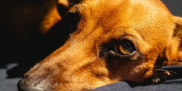
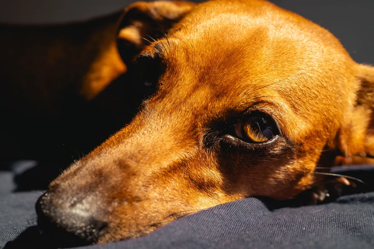
It’s 11 p.m., you’re scrolling through your phone while your usually‑springy Jack Russell Terrier, Milo, lies on the couch with one eye appearing “shrunken” and the lid half‑closed. You notice his nose is a little cooler on that side, too. The sudden change feels alarming, and the word “Horner’s syndrome” you’ve just Googled feels foreign and scary. You wonder: Is this an emergency? Will Milo ever see normally again? And what will the vet bill look like?
We know how unsettling a new, odd‑looking symptom can be. The good news is that Horner’s syndrome itself rarely harms the eye’s vision, but it does point to an issue somewhere along the sympathetic nerve pathway. In this guide we’ll explain what Horner’s syndrome is, why it happens, how to spot it early, what your vet will do to diagnose it, treatment options, expected costs, and how you can help Milo (or any dog) stay comfortable while you work through the diagnosis.
Read on for a step‑by‑step look at the condition, practical tips for home care, and a clear plan for when to call your veterinarian right away.
What is Horner’s syndrome in dogs?
Horner’s syndrome is a collection of signs that result from interruption of the sympathetic nerves that travel from the brain to the eye and surrounding facial structures. In dogs, the classic “3‑S” signs are:
- Ptosis: a drooping upper eyelid.
- Miosis: a constricted (small) pupil.
- Enophthalmos: a sunken appearance of the eye.
Sometimes a fourth sign—heterochromia (different eye colors)—appears if the condition is long‑standing. The syndrome is named after Johann Friedrich Horner, who first described it in humans in the 19th century. In dogs, it’s relatively uncommon but not rare; estimates suggest a few hundred cases per year in the United States, most often seen in middle‑aged to senior dogs of any breed.
What causes Horner’s syndrome?
The cause is usually a disruption of the three‑segment sympathetic pathway:
| Segment | Typical causes |
|---|---|
| First‑order (central) | Brainstem tumor, trauma, or stroke |
| Second‑order (preganglionic) | Neck injury, cervical disc disease, or neoplasia (e.g., lymphoma) |
| Third‑order (postganglionic) | Middle ear disease, otitis media, or a tumor near the sympathetic chain (e.g., parotid gland tumor) |
When the sympathetic nerves are damaged, the opposite parasympathetic signals dominate, producing the characteristic eye changes. The most common primary causes in dogs are:
- Neck or chest trauma (e.g., a car accident).
- Neoplasia, especially nerve‑sheath tumors or lymphoma.
- Middle‑ear disease, such as otitis media with effusion.
- Idiopathic (unknown) causes, which account for roughly 30 % of cases.
Breed predisposition is not strong, but larger breeds with a higher incidence of neck injuries (e.g., Great Danes, Mastiffs) and brachycephalic breeds that are prone to ear infections (e.g., Bulldogs) appear more often in case series.
Signs and symptoms
Owners typically notice the eye changes first, but other subtle clues can appear before the full “3‑S” picture.
| Stage | Typical signs |
|---|---|
| Mild | Just a slightly droopy eyelid or barely noticeable pupil size difference. |
| Moderate | Clear ptosis, noticeable miosis, and mild enophthalmos; the dog may tilt the head to compensate. |
| Severe | Marked drooping, very small pupil, pronounced sunken eye, and possible facial sweating on the affected side. |
Other associated signs can include:
- Cooler skin on the affected side of the face.
- Excessive tearing or nasal discharge if a middle‑ear issue is present.
- Reduced facial muscle tone on the same side.
Vision is usually preserved because the optic nerve (which carries visual information) is not part of the sympathetic pathway. However, if the underlying cause involves the eye itself (rare), vision loss can occur.
When to call your vet
Call your regular vet today if you notice any of the following:
- Drooping eyelid or a pupil that looks smaller than the other eye.
- Visible sunken appearance of the eye.
- Newly cool or sweaty skin on one side of the face.
Go to an emergency veterinary hospital right now if you also see:
- Sudden onset of severe drooping combined with difficulty breathing or coughing.
- Bleeding, obvious trauma to the neck or head, or a collapsing episode.
- Signs of severe pain (whining, inability to settle) that develop rapidly.
These red flags suggest an acute injury or a life‑threatening condition that needs immediate attention.
This article provides general information and is not a substitute for professional veterinary care.
How vets diagnose Horner’s syndrome
Diagnosis is a stepwise process that aims to confirm the syndrome and then uncover the underlying cause.
- History and physical exam: Your vet will ask when the signs started, any recent injuries, and will feel the neck and check the ears.
- Ophthalmic exam: A slit‑lamp or indirect ophthalmoscope helps assess pupil size, lens clarity, and eye pressure.
- Pharmacologic testing: Applying a drop of apraclonidine or phenylephrine can help differentiate between first‑ and third‑order lesions (the pupil will dilate only if the post‑ganglionic nerve is intact).
- Imaging: Depending on the suspected level, your vet may order:
- Radiographs of the neck and chest.
- Advanced imaging (CT or MRI) to look for tumors, disc disease, or ear infections.
- Laboratory work: CBC, chemistry panel, and possibly a fine‑needle aspirate of any mass help rule out systemic disease or cancer.
These tests follow the guidelines from the American College of Veterinary Internal Medicine (ACVIM) and the Merck Veterinary Manual.
Treatment options
Medical treatment
Because Horner’s syndrome itself does not need direct medication, treatment focuses on the underlying cause. Common medical approaches include:
- Antibiotics: For bacterial otitis media, drugs such as amoxicillin‑clavulanate are often used.
- Anti‑inflammatory drugs: Steroids (e.g., prednisolone) may be prescribed to reduce swelling around the nerve.
- Chemotherapy: If a lymphoma is diagnosed, protocols involving vincristine or cyclophosphamide are standard; your oncologist will tailor the plan.
- Pain management: NSAIDs (e.g., carprofen) or gabapentin can help control discomfort.
Ask your vet about these options; dosing will be based on your dog’s weight and specific condition.
Supplements and supportive care
While supplements cannot cure the syndrome, they can aid recovery when the underlying disease involves inflammation or infection:
- Omega‑3 fatty acids (EPA/DHA): Help modulate inflammation, especially in chronic ear disease.
- Probiotics: Support gut health if your dog is on antibiotics.
- Vitamin C: May provide antioxidant support during healing, but discuss with your vet.
Always check with your veterinarian before adding any supplement, as interactions with prescription drugs are possible.
Procedures or surgery
Surgical intervention is considered when a specific structural problem is identified, such as:
- Removal of a middle‑ear mass or drainage of fluid.
- Decompression of a spinal nerve root if a disc herniation is the cause.
- Excision of a peripheral nerve sheath tumor.
Recovery from these procedures typically involves a 1‑ to 2‑week hospital stay, followed by restricted activity for 2‑4 weeks. Costs vary widely; a routine ear surgery may run $1,200–$2,500 USD, while complex spinal surgery can exceed $5,000 USD.
Diet and nutrition
Nutrition doesn’t directly treat Horner’s syndrome, but feeding the right diet can support overall recovery, especially if the underlying cause is an infection, cancer, or chronic inflammation.
| Do feed | Limit | Avoid |
|---|---|---|
| Highly digestible protein sources (e.g., boiled chicken, turkey, or a prescription recovery diet). | High‑fat treats that may exacerbate inflammation. | Raw diets with unknown bacterial load if the dog has an active ear infection. |
| Omega‑3 enriched foods or a fish‑oil supplement (under vet guidance). | Excessive salt, which can worsen hypertension in cancer patients. | Artificial additives or fillers that may irritate a sensitive gut. |
| Small, frequent meals if your dog is recovering from surgery. | Large, single meals that can stress the gastrointestinal tract. | Any diet that your dog has previously shown intolerance to. |
Here are practical feeding tips:
- Transition slowly: If you switch to a prescription or novel protein diet, mix 25 % new food with 75 % old food for the first 2‑3 days, then increase the new portion gradually.
- Hydration matters: Ensure fresh water is always available. For dogs on a high‑protein diet, extra water helps kidney function.
- Monitor weight: Weight loss can indicate an underlying disease; aim for a stable body condition score (BCS) of 4‑5/9.
- Consult a nutritionist: If your dog has a concurrent condition (e.g., renal disease), a board‑certified veterinary nutritionist can tailor a therapeutic diet.
For detailed guidance on calculating caloric needs, you can use our dog nutrition calculator.
Cost and prognosis
Because the cost depends heavily on the underlying cause, it’s useful to break it down into typical ranges (all figures are estimates and can vary by region).
| Component | US estimate | UK estimate |
|---|---|---|
| Initial exam & basic bloodwork | $150–$250 | £80–£150 |
| Imaging (X‑ray) | $200–$350 | £120–£250 |
| Advanced imaging (CT/MRI) | $1,200–$2,500 | £800–£1,600 |
| Middle‑ear surgery | $1,200–$2,500 | £900–£2,000 |
| Chemotherapy (per protocol) | $2,000–$5,000 | £1,500–£4,000 |
| Follow‑up visits (3‑5) | $300–$600 | £150–£300 |
Prognosis varies:
- Idiopathic cases: Often resolve spontaneously within weeks to months; >80 % regain normal eye appearance.
- Traumatic causes: Good to excellent if the nerve isn’t permanently damaged; recovery may take 2–6 weeks.
- Neoplastic causes: Depends on tumor type and stage; some dogs achieve long‑term remission with surgery or chemotherapy, while others have a guarded prognosis.
Overall, most dogs live a normal lifespan after the underlying issue is managed, especially when caught early.
Prevention and home care
Because many triggers are accidental (trauma) or disease‑related, you can’t eliminate all risk, but you can reduce the chance of secondary causes:
- Protect the neck and head: Use a well‑fitted harness instead of a collar for dogs that pull, and avoid rough play that could cause neck injuries.
- Routine ear checks: Inspect the ears weekly for redness, discharge, or foul odor, especially in breeds prone to ear infections.
- Vaccinations and parasite control: Follow AAHA vaccination guidelines and keep fleas/ticks in check to lower the risk of tick‑borne diseases that can affect nerves.
- Regular veterinary wellness exams: Early imaging or blood work can catch hidden tumors before they cause nerve damage.
If your dog is diagnosed, the home care plan often includes:
- Keeping the eye clean with a damp cotton ball (no pressure).
- Administering any prescribed eye drops exactly as directed.
- Limiting vigorous activity for the first few days after surgery or injury.
- Monitoring for changes—new drooping, swelling, or pain—and reporting them promptly.
Vet’s note
From our vet team: “Seeing a droopy eyelid can be frightening, but remember that Horner’s syndrome itself is usually a sign, not the problem. Our priority is finding the root cause—whether it’s an ear infection you can treat with antibiotics, or a more complex tumor that needs imaging. Early detection and a calm, systematic approach give most dogs an excellent chance to recover.”
Key takeaways
- Horner’s syndrome causes a droopy eyelid, small pupil, and sunken eye, but it rarely affects vision.
- Typical causes include neck trauma, ear disease, and tumors; idiopathic cases are also common.
- Prompt veterinary evaluation is essential—call today for any eye changes, and seek emergency care if there’s severe pain or trauma.
- Treatment targets the underlying problem; most dogs improve once the cause is managed.
- Cost varies widely; basic work‑up starts around $150 USD, while advanced imaging or surgery can exceed $2,000 USD.
- Regular ear checks, safe harness use, and routine wellness exams help lower the risk of secondary Horner’s syndrome.
Myth vs. fact
Myth: Horner’s syndrome always means permanent blindness.
Fact: The condition affects the sympathetic nerves, not the optic nerve, so vision is typically preserved.
Myth: It’s a hereditary disease that can’t be prevented.
Fact: While some breeds may be more prone to ear infections that lead to Horner’s, most cases are acquired; good ear hygiene and injury prevention can reduce risk.
Myth: Surgery is required for every dog with Horner’s syndrome.
Fact: Surgery is only needed when a specific structural problem (e.g., a tumor or ear mass) is identified; many cases resolve with medical management alone.
Frequently asked questions
What causes Horner’s syndrome in dogs?
Most often it results from neck or chest trauma, ear infections, or tumors that compress the sympathetic nerve pathway; sometimes the cause remains unknown.
Why does my dog have a droopy eyelid and small pupil?
These are the hallmark signs of Horner’s syndrome, indicating a disruption of the sympathetic nerves that control eye muscles and pupil size.
Is Horner’s syndrome hereditary?
There’s no strong genetic link, though breeds prone to ear disease (e.g., Bulldogs) may see it more frequently due to secondary infections.
Will my dog lose vision with Horner’s syndrome?
Vision is usually unaffected because the optic nerve isn’t part of the sympathetic pathway; however, underlying diseases could threaten eyesight if left untreated.
How long does recovery take after treatment?
Recovery depends on the underlying cause—idiopathic cases may improve in a few weeks, while surgical or oncologic cases can require months of follow‑up.
Do I need surgery for Horner’s syndrome?
Surgery is only indicated when a specific structural problem (like a tumor or ear mass) is identified; many dogs recover with medication and supportive care alone.
Ask the PuppaDogs community
Have a question this article didn’t fully answer? Want to compare notes with other dog owners who’ve been through this? Our community forum is moderated by experienced owners and vets — and answers tend to come fast. Ask in the PuppaDogs community →
References
- American College of Veterinary Internal Medicine (ACVIM) – Neurology Guidelines, 2022.
- Merck Veterinary Manual, “Horner’s Syndrome” entry, 2023 edition.
- American Animal Hospital Association (AAHA) – Recommended Diagnostic Imaging Protocols, 2021.
- Veterinary Ophthalmology, “Sympathetic Nerve Pathways in Dogs,” 2020.
- University of California, Davis Veterinary Medicine – Otitis Media and Neurologic Complications, 2022.
- World Small Animal Veterinary Association (WSAVA) – Pain Management Standards, 2023.


